|
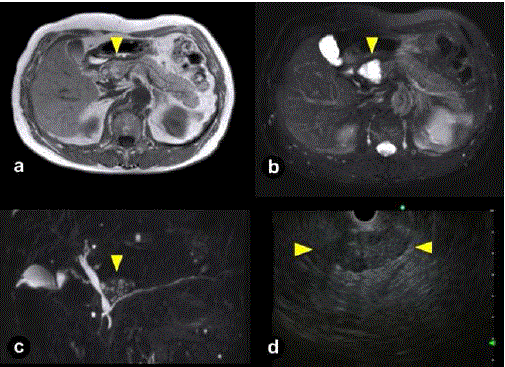
| Figure 2. Magnetic resonance imaging (a., b., c.) and endoscopic ultrasonography (d.) of LECs with sebaceous glands in 2013. a. T1-weighted image revealed hypo- and hyper-intensity (yellow arrow head). b. T2-weighted image identified a hyper-intense polycystic lesion which protruded toward extra-pancreas (yellow arrow head). c. MRCP also revealed a polycystic lesion which resembled cheerios-like appearance (yellow arrow head). d. EUS revealed a solid-appearing lesion, measuring 4 cm, with slight posterior enhancement in the pancreatic head (between yellow arrow heads). |