|
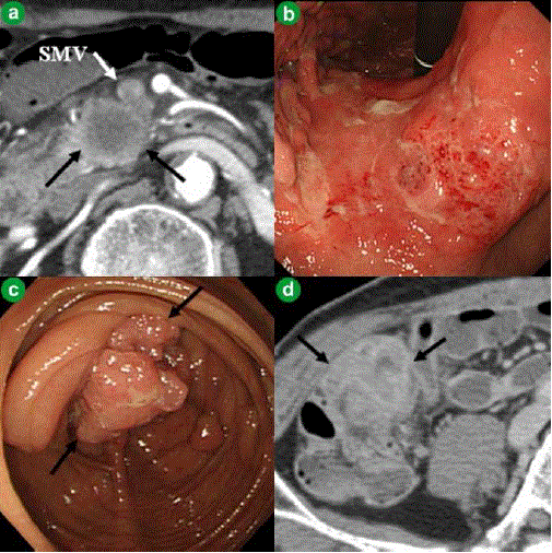
| Figure 1. Appearance of the three cancers at diagnosis. a. Enhanced computed tomography showing a pancreatic head tumor 22 mm in diameter (black arrows) with superior mesenteric vein (SMV) invasion (white arrows). b. Upper gastrointestinal endoscopy showing a gastric tumor in the middle part of the stomach. c. Lower gastrointestinal endoscopy showing cecal cancer (black arrows). d. Computed tomography showing a large cecal tumor (black arrows). |