|
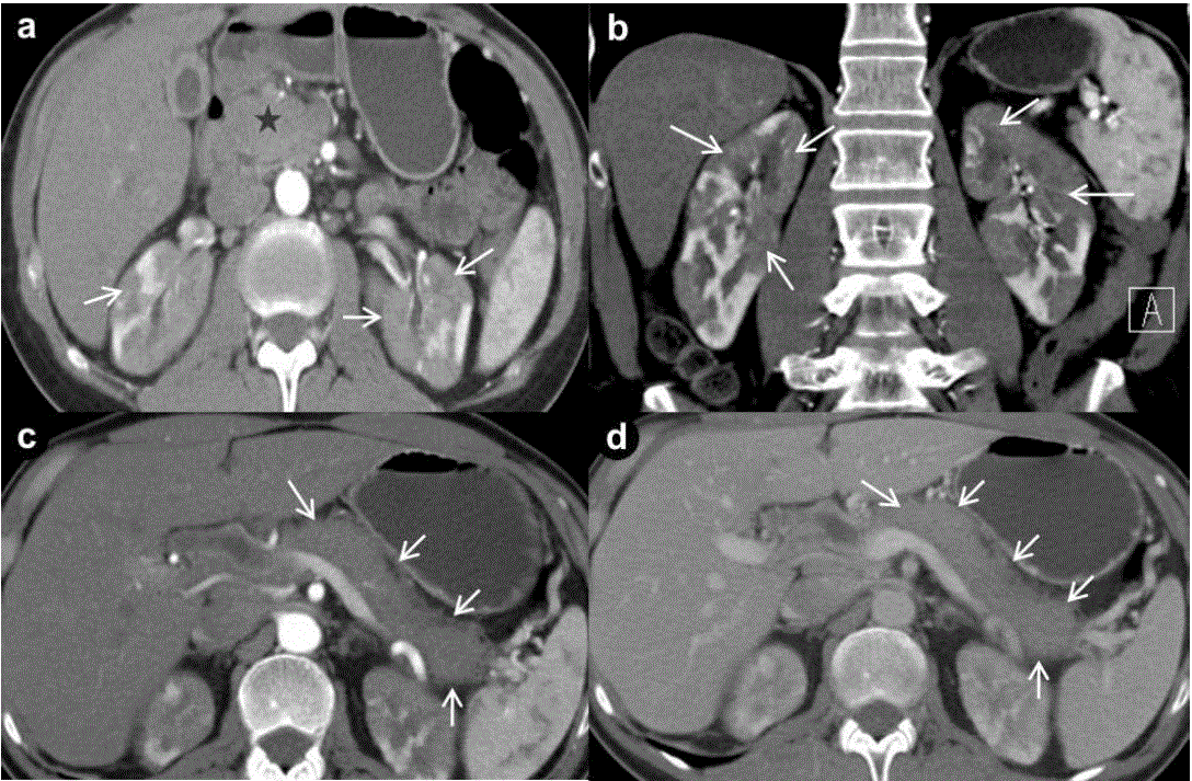
| Figure 1. Pre treatment axial (a.) and coronal (b.) CT demonstrates enlarged pancreas and patchy areas of decreased enhancement in bilateral renal parenchyma (arrows). Diffusely enlarged pancreas (arrows) demonstrating the body and tail in arterial phase (c.) and in venous phase (d.). |