|
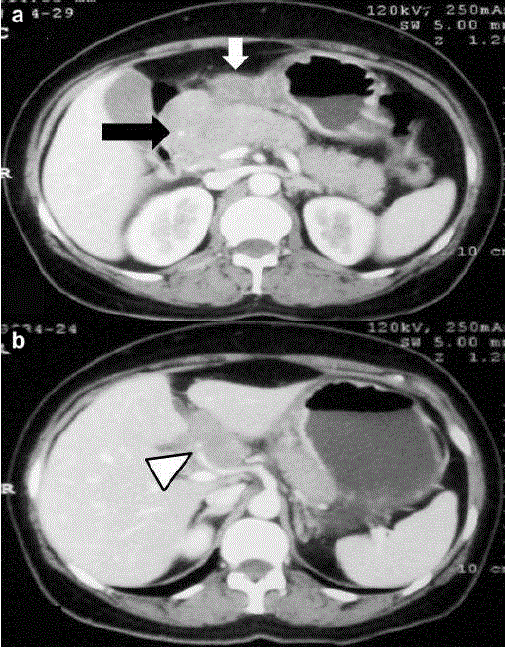
| Figure 1. Contrast CT abdomen showing (a.) thickening of gastric body and antrum (white block arrow) along with conglomerated lymph node mass in the peripancreatic region (black block arrow) with clear fat plane between the mass and pancreatic head and (b.) hepatic artery encasement (white arrow head). |