 |
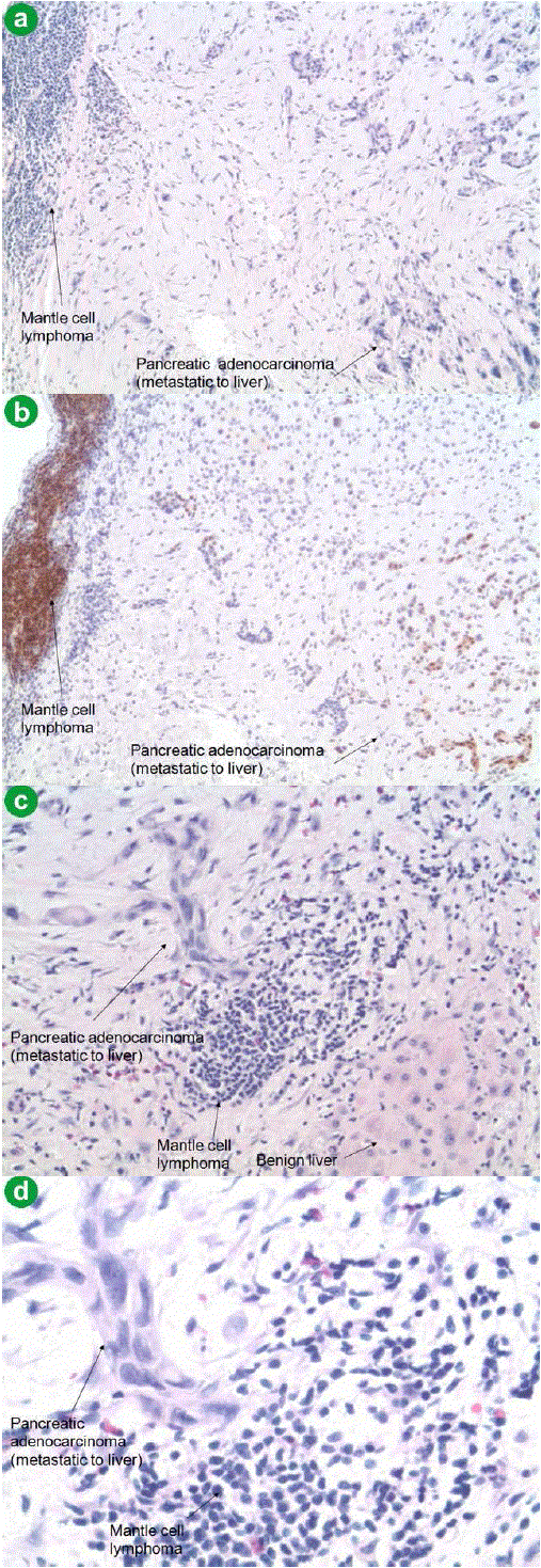
| Figure 4. Pathology microphotographs of the liver core biopsy. a. Liver biopsy specimen showing a diffuse monomorphic infiltrate of smallto- medium sized lymphocytes and adjacent metastatic poorly differentiated pancreatic adenocarcinoma (H&E, 4x magnification). b. Mantle cell lymphoma showing positive staining for cyclin D1 immunohistochemical stain (H&E, 4x magnification). c. Liver biopsy showing a diffuse monomorphic infiltrate of small-to-medium sized lymphocytes and adjacent metastatic poorly differentiated pancreatic adenocarcinoma (H&E, 20x magnification). d. Liver biopsy showing a diffuse monomorphic infiltrate of small-to-medium sized lymphocytes and adjacent metastatic poorly differentiated pancreatic adenocarcinoma (H&E, 40x magnification). |