 |
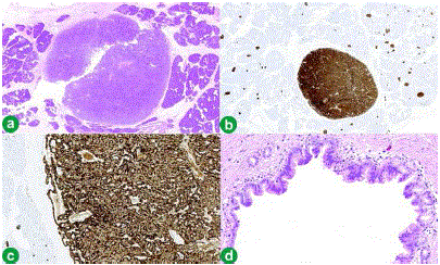
| Figure 4. a. Well-circumscribed tumour mass surrounded by pancreatic tissue (haematoxylin and eosin stain x1.25). b. Diffuse staining of tumour and positive staining of islets of Langerhans in surrounding tissue (immunohistochemistry: chromogranin A stain x1.25). c. Higher power image showing positive staining of tumour and islets of Langerhans in surrounding tissue (immunohistochemistry: chromogranin A stain x10). d. IPMN showing papillary formation (haematoxylin and eosin stain x20). |