|
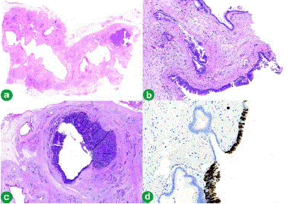
| Figure 3. a. Mega block showing cystic lesions of IPMN (haematoxylin and eosin stain x1). b. IPMN showing neoplastic epithelium with micropapillary formation and overlapping nuclei indicating high-grade dysplasia (haematoxylin and eosin stain x10). c. Early invasive neuroendocrine carcinoma and neuroendocrine carcinoma in situ with cystic change in the tail of the pancreas (haematoxylin and eosin stain x1.25). d) IPMN (pancreatic subtype) showing strong positive staining (immunohistochemistry: Muc5AC stain x10). |