|
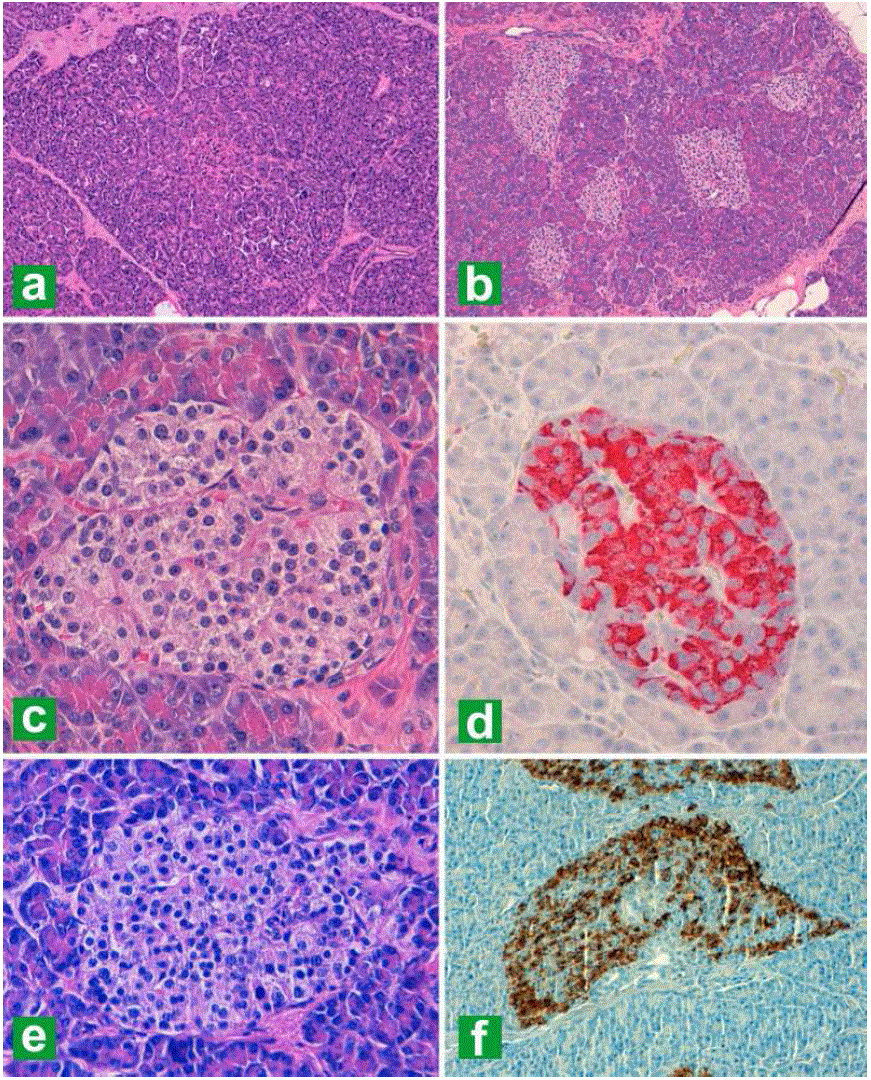
| Figure 1. a. Control pancreatic specimen (hematoxylin-eosin stain (H&E), original magnification 100x). b. Pancreatic tissue derived from the first pancreatic resection in 2001 showing increased and irregular-shaped islets (H&E, original magnification 100x). c. Islet-cell hyperplasia with slight variations in nuclear size (H&E, original magnification 400x). d. Langerhans islet with predominance of insulin positive cells (anti-insulin 1:100, original magnification 400x). H&E (e.) and anti-insulin stain (f.) of pancreatic tissue derived from the resection of the remaining pancreatic tissue in 2012 (original magnification 400x for both). |