 |
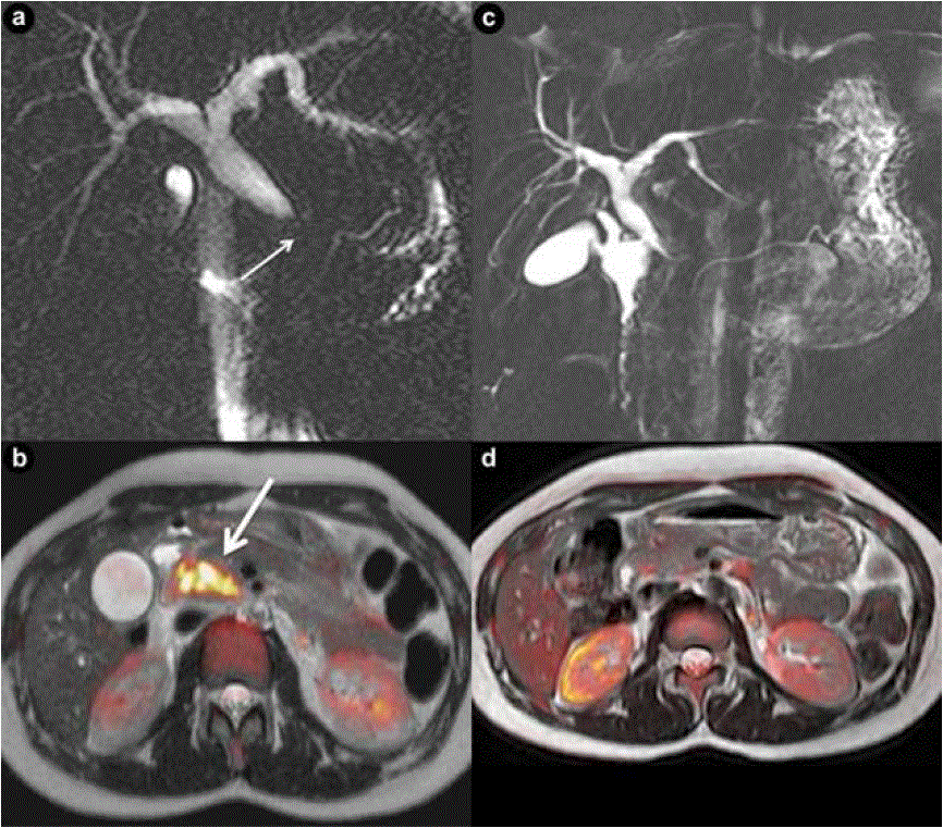
| Figure 3. a. MRCP shows distal common bile duct obstruction (short arrow). Main pancreatic duct is not dilated. b. Coloured map of T2-weighted and Diffusion-weighted sections through the head of the pancreas displaying a space-occupying lesion with cellular proliferation (long arrow). Control MRI shows almost complete resolution of bile duct obstruction (c.) and a normal pancreatic head (d.). (Patient #3). |