|
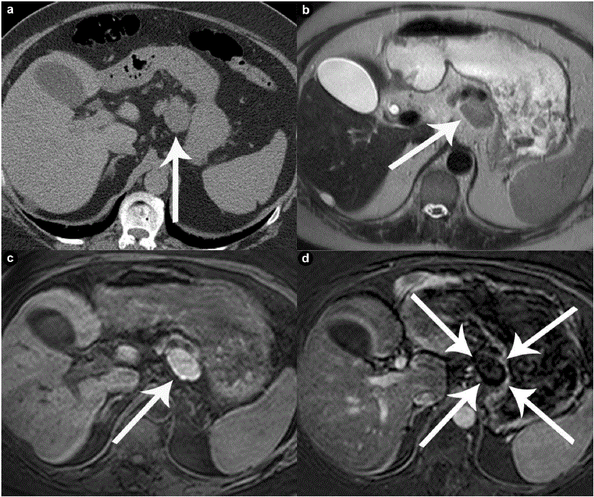
| Figure 3. A 63-year-old male with incidentally discovered pancreatic mass (Case #5). a. Unenhanced axial CT shows a well defined mass arising off the superior aspect of the pancreatic body (arrow) measuring above fluid attenuation, and incompletely evaluated without intravenous contrast. Due to decreased glomerular filtration rate, MRI was used to characterize the lesion. Axial T2 weighted images (b.) show a well defined mass of intermediate signal intensity, which shows inherent high signal intensity on unenhanced T1 weighted images (c.). T1 post-contrast subtraction imaging (d.) shows the mass having a black non-enhancing center, with a thin rim of peripheral enhancement. |