|
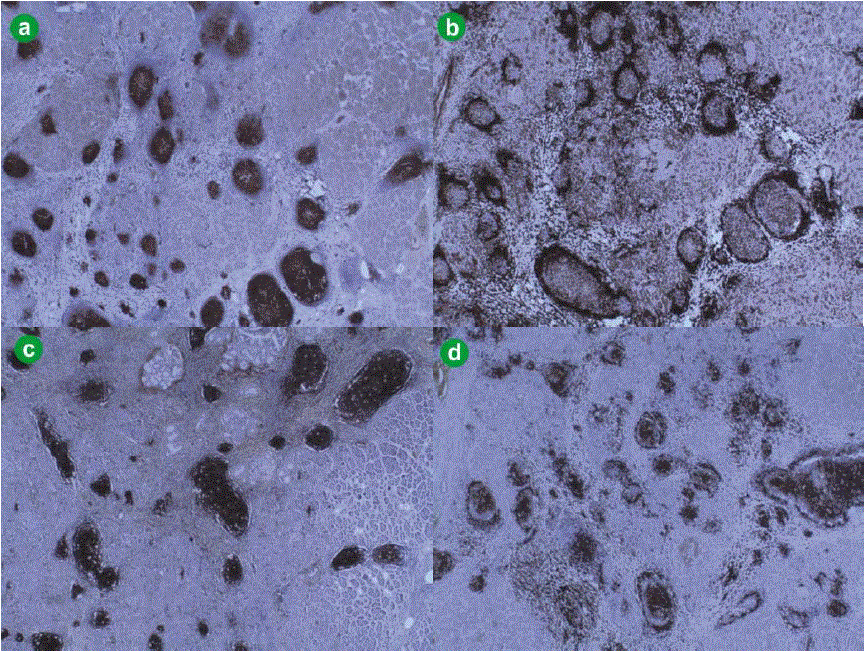
| Figure 2. Immunohistochemical staining revealed a follicular infiltrate which was composed of centers with cells expressing B-cell characteristicsthat were CD10 positive (a,) and BCL-2 negative (b.) (BCL-2 negativity implies reactive rather than neoplastic follicles), whilst CD21 (c.) and CD23 (d.) staining demonstrated regular and orderly network of follicular dendritic cells as in reactive lymphoid follicles, thus supporting the diagnosis of reactive lymphoid follicular hyperplasia. |