|
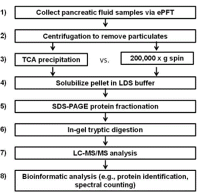
| Figure 1. Experimental workflow. 1) Pancreatic fluid was collected via ePFT. 2) Samples were centrifuged to remove any particulates. 3) Pancreatic fluid was precipitated with TCA or subjected to ultracentrifugation at 200,000 g for 2 h. 4) The pellet was solubilized in LDS Laemmli loading buffer. 5) Proteins were fractionated by SDS-PAGE. 6) Proteins were in-gel typically digested. 7) Resulting peptides were subjected to LC-MS/MS analysis. 8) Proteins were identified via database searching using Proteome Discoverer (Thermo Scientific, Waltham, MA, USA) and Scaffold 3 (Proteome Software Inc., Portland, OR, USA) software. |