|
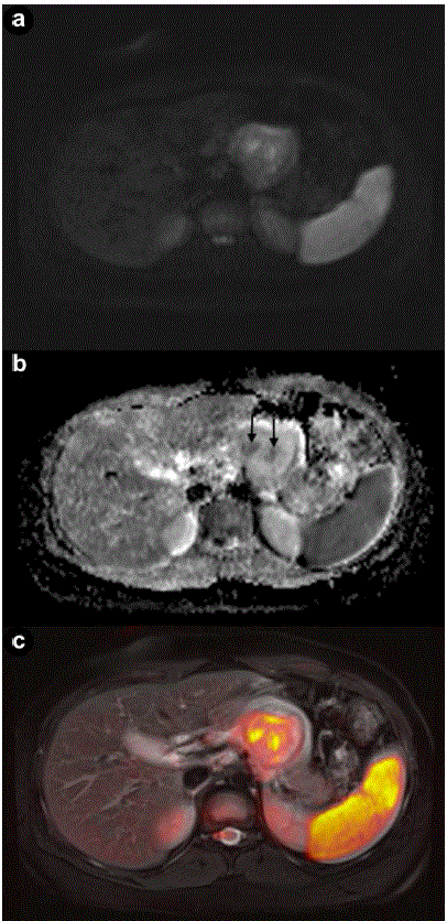
| Figure 4. a. Axial diffusion-weighted image (b value of 800 sec/mm2) shows a lesion with low signal intensity with some small central areas of higher signal intensity from restricted diffusion. b. Apparent diffusion coefficient mean value of the lesion is 1.8x10⁻³ mm2/sec with the central hypointense areas having a minimum value of 1.3x10⁻³ mm2/sec (arrows). c. T2-weighted and diffusion-weighted imaging were fused displaying a colored map of abnormal diffusion parameters. |