|
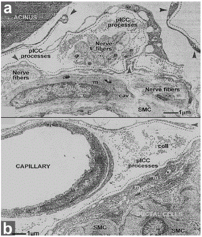
| Figure 6. Transmission electron microscopic images from rat exocrine pancreas showing the spatial relationship of the pancreatic interstitial cells of Cajal and their processes with acini, unmyelinated terminal nerve fibers, arteriolar smooth muscle cells (a.), capillaries or ductal cells (b.). The cytoplasmic processes of pancreatic interstitial cells of Cajal (black dashed lines) can be easily recognized: very long, thin and with dilatations (“moniliform” aspect), containing mitochondria and other organelles (arrowheads). (Reproduced with permission from Popescu et al. [25]). cav: caveolae; col: collagen; m: mitochondria; E: endothelium; SMC: smooth muscle cells |