|
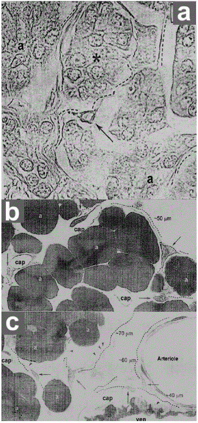
| Figure 5. a. Conventional paraffin embedded light microscopic tissue sections of the human exocrine pancreas (methylene blue staining, 40x). In the interstitium, amongst the acini (a), note some spindle-shaped or triangular cells (arrows), with very long cytoplasmic processes (several tens of μm), made evidently by black dashed lines. Such cells could be interstitial Cajal-like cells, actually pancreatic interstitial cells of Cajal. The acini marked by asterisks appear surrounded by periacinar pancreatic interstitial cells of Cajal processes. (Reproduced with permission from Popescu et al. [25]). b. c. Non-conventional semi thin epon-embedded tissue sections from rat exocrine pancreas stained with toludine blue. Numerous pancreatic interstitial cells of Cajal are present in the interstitium in between the acini (about 80). Note that only the pancreatic interstitial cells of Cajal body (arrows) and the emerging very long (20-50-100 μm), thin (less than 0.5 μm) cytoplasmic processes (black dashed lines) with characteristic “moniliform” aspect (arrowheads) are evident. (400x). (Reproduced with permission from Popescu et al. [25]). a: acini; cap: capillary; art: arteriole; ven: venule |