|
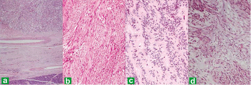
| Figure 3. Photomicrographs from the tumor in the pancreas showing pancreatic tissue at the periphery and the cellular lesion (40x) (a.). The tumorshowed predominantly fusiform spindle shaped cells in intersecting fascicles resembling a smooth muscle tumor (b.), with foci of nuclear pallisading reminiscent of a neural tumor (c.), and myxoid areas (d.) (H&E, 100x). |