|
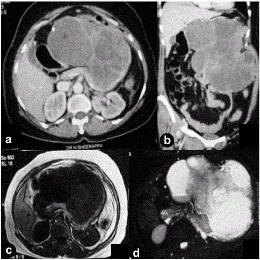
| Figure 1. Contrast-enhanced computerized tomogram of the abdomen: axial (a.) and coronal (b.) sections showing a large lobulated heterogeneously enhancing mixed echogenic lesion in the region of the body and the tail of the pancreas. On magnetic resonance imaging, the mass was hypointense on T1-weighted imaging (c.) and hyperintense on T2- weighted imaging (d.). |