|
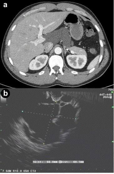
| Figure 2. a. Contrast enhanced abdominal CT scan showing a cystic lesion adjacent to the body of the pancreas and extending into the porta hepatis from Case #2. b. Linear EUS image showing the anechoic cyst with several thin septae from the same patient. |