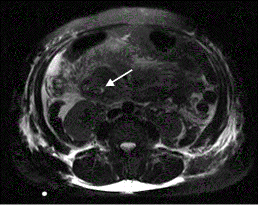
Figure 2.
Axial MRCP (T2 HASTE sequence, thin slice) image demonstrating intraluminal filling defect due to ampullary lesion (white arrow).