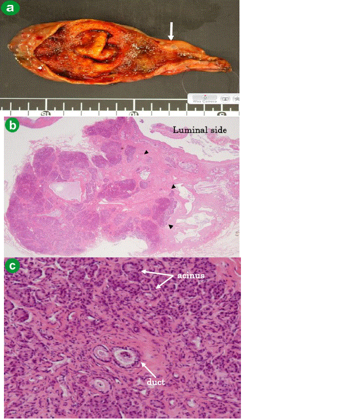
Figure 3.
Image of protruding lesion near the cystic duct (a. arrow). The microscopy reveals pancreatic acinar and duct content in the layer of subserosa (b. H&E, x100; c. H&E, x400).