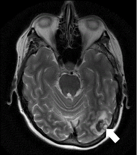
Figure 3.
T-2 weighted magnetic resonance imaging demonstrating a 1.5 cm area of intraparenchymal hemorrhage in the posterior left temporal lobe (white arrow).