|
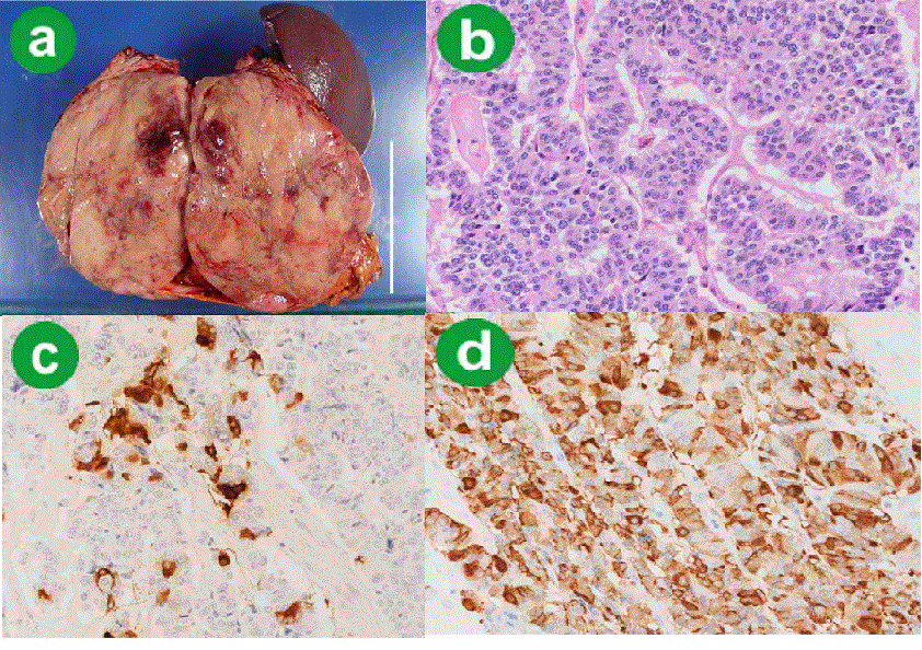
| Figure 2.The macroscopic and histological appearance of the resected tumor. a. The resected tumor was solid, and there was a small focus of hemorrhage on the cut surface (scale equal to 10 cm). b. The tumor cells had lightly eosinophilic cytoplasm and proliferated around the blood vessels. c. Approximately 10% of tumor cells were immunostained with VIP. d. The most of the tumor cells were positive for calcitonin. |