|
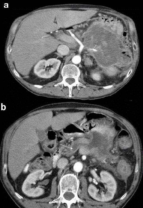
| Figure 1.Preoperative contrast-enhanced CT-scan at the celiac axis level. a. A large low-density mass in the pancreatic body and tail infiltrating the posterior gastric wall, the splenic flexure of the colon and the spleen. The amputation of the splenic artery is evident. b. Dilated main pancreatic duct (white arrow). |