|
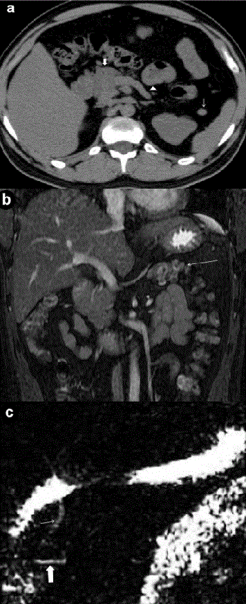
| Figure 1. Case #1. a. Axial non-contrast computed tomography image shows normal size pancreatic head (thick arrow), splenunculus (thin arrow), stomach and bowel loops in pancreatic bed anterior to splenic vein(arrow head) described as dependent stomach and dependent intestine signs. b. Coronal T2W fast imaging employing steady state acquisition MR image depicts transverse colon in distal pancreatic bed (arrow). c. MRCP image demonstrates ventral pancreatic duct (thick arrow) and common bile duct (thin arrow). |