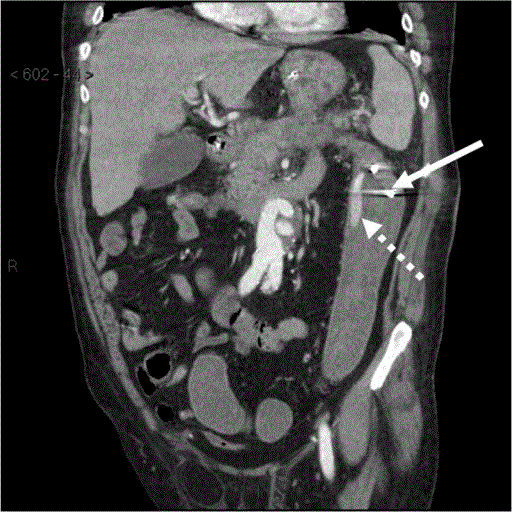
Figure 2.
Coronal reformatted CT image demonstrates the position of the percutaneous drain in the colon lumen (arrow) and extravasated intravenous contrast mixed with unopacified blood in the descending colon (interrupted arrow).