 |
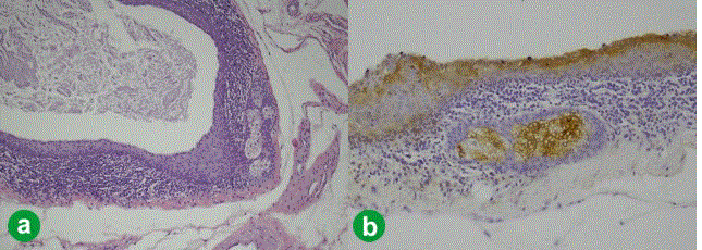
| Figure 4.a. Microscopic analysis revealing an intrapancreatic cystic neoplasm with stratified keratinized squamous epithelium and sebaceous glands, surrounded by a wall of lymphoid tissue (hematoxylin and eosin staining 10x). b. Immunohistochemical analysis of epithelial membrane antigen (EMA) expression in the resected pancreatic tissue, showing the staining of sebaceous glands (anti-human EMA, E29 clone, DakoCytomation, Glostrup, Denmark; 10x). |