 |
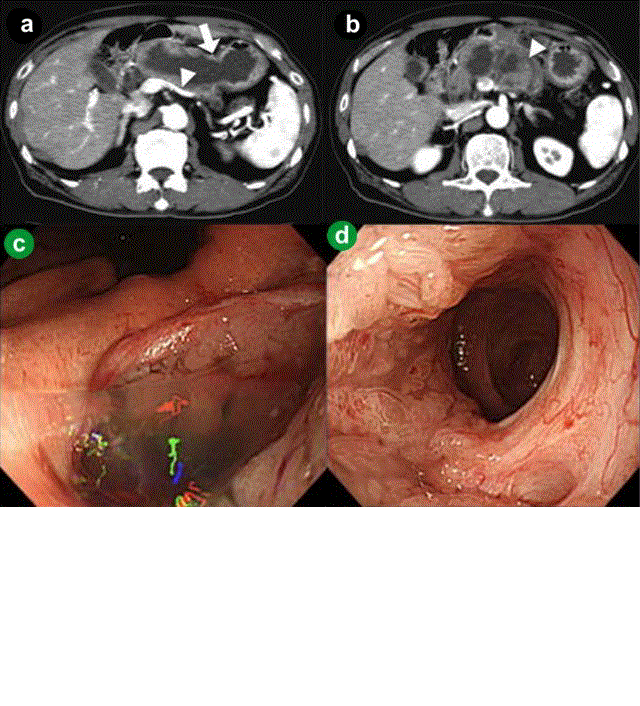
| Figure 3.a. Computed tomography (CT) showed the main pancreatic duct penetrating to the stomach (arrow) and reperfusion of the splenic vein (arrowhead). b. CT showed increased size of the papillary tumor in the main pancreatic duct. c. Gastroduodenoscopy showed gastropancreatic fistula on the lesser curvature of the middle gastric body. Mucus was discharged from the fistula. d. Suction of the mucus enabled us to pass an endoscope into the dilated main pancreatic duct through the fistula and directly examine the lumen of the main pancreatic duct and the papillary tumor adjacent to the fistula. |