|
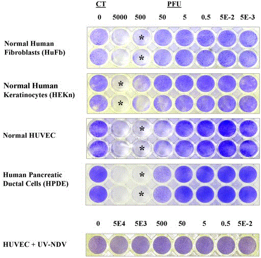
| Figure 5.Photographs of multiwell plates stained with crystal violet showing NDV-LS cytotoxicity in normal human cell lines. In duplicate rows of wells, unstained wells represent those in which total cytotoxicity and lysis had occurred. The far left well in each row was a negative control that received no NDV. Asterisks (*) mark the wells where the minimum cytotoxic dose of NDV was seen for HuFbs, HEKn, HUVEC, HPDE cultures. Acetylated trypsin had been present in all wells shown except those containing HEKn cells. Even at 50,000 PFU, UV-inactivated NDV-LS had little effect on HUVEC cell viability. |