 |
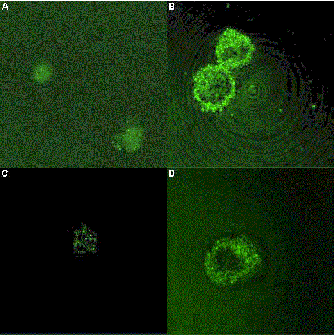
| Figure 3: Cytoplasmic CCR3 and CCR5 staining as assessed by confocal microscopy in control and IL-1β- treated RIN-5AH cells: A) cytoplasmic CCR3 without IL-1β; B) cytoplasmic CCR3 with IL-1β; C) cytoplasmic CCR5 without IL-1β; D) cytoplasmic CCR5 with IL-1β. |