|
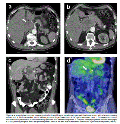
| Figure 1. a. Arterial phase computed tomography showing in axial image a partially cystic pancreatic head mass (arrow) with celiac artery running adjacent to it. b. The mass extended into the uncinate process of the pancreas adjacent to the superior mesenteric artery. c. The mass seen on coronal reconstruction in porto-venous phase demonstrating cystic and solid (asterisk) components. d. Computer tomography positron emission tomography (CT-PET) showing no uptake within the cystic component (arrow) of the mass with mild increased uptake in the adjacent solid component (asterisk). |