|
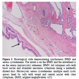
| Figure 3. Histological slide demonstrating synchronous IPMN and serous cystadenoma. The arrow is on the IPMN and the arrowhead is on the serous (microcystic) adenoma. IPMN: tall columnar cells with basal nuclei and abundant mucinous cytoplasm lining a markedly dilated duct. Microcystic serous cystadenoma: multiple small cystic spaces lined by cells with round and central nuclei with clear cytoplasm. (H&E; original magnification: x25) |