Neurosciences & Brain Imaging Open Access
-
Journal h-index: 1
- Average acceptance to publication time (5-7 days)
-
Average article processing time (30-45 days)
Less than 5 volumes 30 days
8 - 9 volumes 40 days
10 and more volumes 45 days
Editorial - (2017) Volume 1, Issue 1
Angel Molina Leon*
Neurophysiology Clinical Center Virgen De La Caridad Cartagena-Murcia Spain
*Corresponding Author:
Angel Molina Leon
Neurophysiology Clinical Center Virgen De
La Caridad Cartagena-Murcia Spain
Tel: + 968325258
E-mail: molinadelaasuncion@gmail.com
Received date: September 28, 2017; Accepted date: September 28, 2017; Published date: September 30, 2017
Citation: Leon AM (2017) Electromyography. Neurosci Brain Imaging. Vol. 1 No. 1:2.
Copyright: © 2017 Leon AM, et al. This is an open-access article distributed under the terms of the Creative Commons Attribution License, which permits unrestricted use, distribution, and reproduction in any medium, provided the original author and source are credited.
Editorial
What is Electromyography and what is used
Electromyography (EMG) is the electrophysiological study of the neuromuscular system. It is a complementary test, and prolongation of the clinical neurological study. Given the broad neuromuscular system and brain, in Spain there is a specific and specific pathway via MIR of Neuromuscular which is called Clinical Neurophysiology which includes EEG, EMG, Multimodal Potentials (PEAT, PEV, PESS) and Intraoperative Monitoring. In addition to sleep pathology, and polysomnography and is taught by physicians via MIR with 4 years of training within this specialty. The EMG is designed in each case according to each protocol, according to the specific pathology, it is not the same to see a syndroem of the carpal tunnel that to see a Polyneuropathy or to study in depth a patient with suspicion of lateral sclerosis amiotrofica ELA, and in function of this each protocol has a different duration. One of the characteristics in my judgment of a good neurophysiologist is meticulousness, patience, the taste for this part of medicine and objectivity, it is very important to be objective [1-3].
The test consists of two part electroneurography and electromyography (Figure 1).
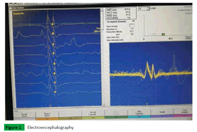
Figure 1: Electroencephalography.
Indications of electromyography
• Differentiation between weakness of central or peripheral origin.
• Differentiation between weakness of neurogenic or myogenic origin.
• Differentiation between preganglionic (radicular) or postganglionic (plexular/truncal) lesion.
• Location of the lesion in the mononeuropathies compressive or traumatic and determination of the degree of affectation (focal demyelination versus axonal degeneration) [4,5].
• Differentiation between multifocal neuropathies and polyneuropathies; degree of involvement of the motor and sensory fibers.
• Differentiation between demyelinating and axonal neuropathies.
• Determination of prognosis in neuropathies.
• Characterization of neuromuscular junction disorders (pre- or postsynaptic).
• Identification of signs of denervation, fasciculations, myotonia and neuromyotonia in "normal" muscles.
• Differentiation between cramp and contracture [6].
Electroneurography
Consists of the study of nerves, this sensory conduction, motor conduction, F waves, reflex blink reflex, H reflex, repetitive stimulation, pudendal nerve protocol, etc. This technique is based on the electrical stimulation of a nerve and the recording of the potential caused by placing electrodes to measure it well in the same nerve, or in the muscle innervated by it. The potential is, therefore, and unlike those spontaneously generated by brain and muscle, an artificially produced potential. Latency, conduction velocity (the time it takes for the potential to occur) to be evaluated; the amplitude, that is to say, the functioning volume of structures that generate the response, and the form thereof, i.e. the functional integration of said structure. Latency expresses the degree of myelination of nerve fibers. Increased latency, or even a decrease in nerve conduction velocity, are signs of demyelination [7].
The amplitude of the potential accounts for the number of response generators. At a potential recorded on a nerve, the decrease in amplitude is a sign of axon loss. At the potential recorded in a muscle the decrease in amplitude may mean either axons lost, or the inability of the muscle to respond.
Electromiography study of muscle functionality, there are three types of normal, neuropathic and or myopathic patterns.
Importance of EMG in different syndromes
Mononeuropathies and entrapment syndromes:
Pathophysiology of traumatic nerve injuries (neuroapraxia, axonotmesis neurotmesis)
Clinical presentation of traumatic injuries:
• In acute injuries
• Acute compression injuries
• Chronic compression injuries - Actrapamientos.
EMG diagnosis, classification and prognosis.
Electrophysiological studies in the reinnervation, aberrant axonal regeneration.
Plexopathies and Radiculopathies
Brachial plexopathy and lumbosacral: Idiopathic, traumatic, postradiation
Pathophysiology of Radiculopathies
Specific clinical situations
Electrophysiological alterations in radiculopathies. Regional Considerations
Value and Limitations of Electrophysiological Study
Intraoperative monitoring
Diseases of motor neurons and spinal cord
Amyotrophic lateral sclerosis, clinical considerations and electrophysiological diagnosis and involvement in the differential diagnosis with motor neuronopathy with anti-GM1 antibodies
Implications of EMG in the diagnosis and assessment of: Poliomyelitis-Post-polio syndrome; viral infections of motor neurons [8].
2nd focal neuron syndromes: Mononmyelic amyotrophy, Syringomyelia
Congenital and hereditary neuropathies
Clinical pathophysiology and etiopathogenic advances (molecular genetic studies) of neuropathies whose primary polyneurology is the primary or predominant: hereditary sensory-motor neuropathy, neuropathy due to excessive pressure vulnerability, familial amyloidotic polyneuropathies, hereditary sensory and dysautonomic neuropathies, hereditary motor neuronopathies, bulb-spinal neuronopathy linked to the X chromosome. Electrophysiological studies based on diagnosis. Classification and nosological characterization.Neuropathies whose polyneuritic syndrome is accompanied by semiologic by affectation of other systems (neurological or non-neurological) [9].
Acquired neuropathies
Physiology: Pathophysiology of conduction block, demyelination and axonal degeneration, electroneurographic quantification. Electrophysiological studies in different dysimmune neuropathies (AIDP, CIDP, multifocal motor neuropathy with conduction blockages) performance and limitations in diagnosis, subtype differentiation and prognosis.
Characterization of the different forms of diabetic neuropathy.
Differential features of EMG results in different toxic and metabolic neuropathies.
Neuromuscular transmission disorders.
Pathophysiology of neuromuscular transmission.
Electrophysiological study in postsynaptic origin processes (myasthenia gravis) its diagnostic performance in the different forms.
Study in processes of presynaptic origin (Eaton Lambert syndrome, Botulinum intoxication).
Clinical and electrophysiological characterization of the main congenital myasthenic syndromes.
Myopathies
Clinical and pathophysiology of genetic processes (Dystrophinopathies sarcoglicanopathies, channelopathies) and acquired (inflammatory, toxic, endocrine myopathies) that affect muscular function.
Development and application of quantitative EMG techniques, correlation and support to the indication of histological studies and their limitations.
Cranial neuropathy
Assessment and prognosis in facial paralysis.
Hemifacial spasm.
Syndromes with motor axonal hyperactivity
Central Nervous System:
Electrophysiological study in movement disorders (Tremor, myoclonus, cranio-cervical dystonia and spasm of the clerk ...).
Multisystemic atrophy.
