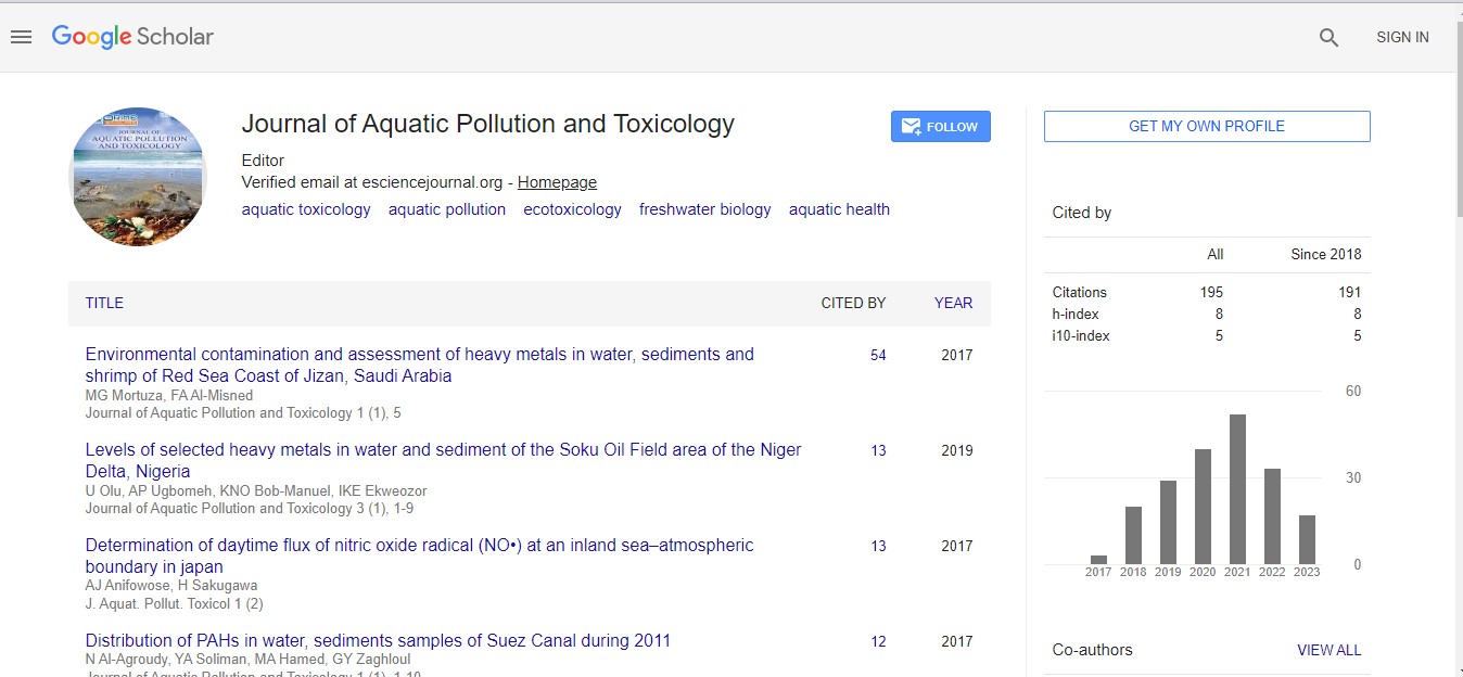
Keywords
Callyspongia fibrosa; Fasciospongia cavernosa; Dysidia fragilis; Potential toxic elements; Tuticorin
Introduction
Gulf of Mannar (GoM) was declared as the first marine biosphere reserve of India and of the South East Asia in the year 1989 [1, 2]. The GoM has a coast line of approximately 10, 800 Sq. Km and consists of 21 uninhabited islands beginning from the holy town of Rameswaram to the industrial town of Tuticorin. It consists of diverse ecosystem constituting a number of flora and fauna especially dominated by corals and marine sponges [1-3]. Tuticorin is industrial city which is heavy threatened by pollutants from industries which usually include chemical processing industries, alkali plants, petrochemical industries, thermal power plant and sewage discharge from the local municipalities [2, 4]. The mangrove vegetation which is most prominent in Tuticorin is facing the threat of extinction due to the impact of the effluents released into the coast. Among all these industries, contribution from thermal power plant is seen causing significant enrichment of PTEs in the marine sediments of Tuticorin [2, 5]. The fly ash and slurry discharged from these units also resulted in the significant enrichment of PTEs in the coast and resulted in the irreversible damage to the ecosystem [2]. The increased release of the contaminants from the surrounding industry into the coast may play a substantial role on PTEs pollution.
The pollution caused by PTEs has been studied in different coastal ecosystems of the world [6]. This is one of the major environmental problems being faced by a number of coastal ecosystems during the recent past due to rapid industrialization and anthropogenic activity posing a serious risk to the inhabiting aquatic biota. PTEs emitted from agriculture, industries and domestic sources have an impending effecting on the existing biota [2]. The concentration of the PTEs in seawater and sediments increases due to excessive release of effluents both from domestic and industrial sources [2]. Among the environmental contaminants PTEs are seen posing major ecological consequences [5]. A number of biota had been used to monitor the concentration of PTEs in different coastal regions of the world but still the most preferred are the marine invertebrates [7]. The use of mussels in “Mussel Watch programmer” in North America to monitor the concentration of PTEs in coastal environments had given good results but with some limitations [8]. However, other researchers have emphasised the need to use other filter feeding organisms such as barnacles, polychaetes, tunicates and the sponges which are sedentary, filter feeding and sustain the fluctuation in environment have also been used as bio monitors for PTEs contamination [1, 2, 7-14]. Sponges are considered as good bio monitors by a number of researchers because of their unique features and their adaptations to a number of ecological niches [15]. The long life span, adherent natures, survival under extreme physical and chemical fluctuations, accumulation of contaminants both from dissolved and suspended phases all these features makes sponges an ideal bio monitor [1, 2, 7, 8, 14, 16]. PTEs concentration in the fish clams and gastropods may show significant difference [17]. In contrast, the sessile and benthic organism such as sponges significantly reduces this variation in the concentration of PTEs. This affirmation is based on the assumption that variation in PTEs concentration within the different sponge species of the same area are lacking? Here we measured the concentration of As, Cd, Co, Cr, Cu, Fe, Ni, Pb and Zn in C. fibrosa, F. cavernosa and D. fragilis collected from Tuticorin coast. The concentration of nine PTEs in three species was compared at five stations (S1-S5) along the coast of Tuticorin.
Materials and Methods
Sample collection and identification
Marine sponges Callyspongia fibrosa (Ridley & Dendy, 1886, Family: Callyspongiidae, Order: Haplosclerida), Faciospongia cavernosa (Schmidt, 1862; Family: Thorectidae) and Dysidia fragilis (Family: Dysideidae, Order: Dictyoceratida) were collected along the coastal regions of Tuticorin, India. Sponge specimens were collected by snorkelling and skin diving from five different stations between 8o 30’ and 8o 46’ N latitude 78o 08’ and 78o 15’ E longitude in the state of Tamil Nadu, India and their morphological description is presented in Table 1 and Figure 1. The specimens were cut using a ceramic knife to avoid PTEs contamination, these samples were stored in zip lock bags and later washed thoroughly with natural seawater and the foreign matter such as mud particles, macro detritus and other organisms which are found in the sponges were removed by monitoring under the stereomicroscope with the help of plastic tweezers. The voucher specimens were submitted to National Institute of Oceanography (NIO) Goa, for depository purpose and were identified by Dr. Thomas as C. fibrosa, F. cavernosa and D. fragilis, at Central Marine Fisheries Research Institute (ICAR), Vizhinjam, Thiruvananthapuram, India.
Sample preparation
The oven dried specimen samples of D. fragilis, C. fibrosa and F. cavernosa were used for the analysis of PTEs by the method of Mc Carthy and Ellis [18]. The samples were placed in Teflon beakers and digested with 10 ml of 70% nitric acid (HNO3) in a microwave vessel in an IFB (Indian Fine Blanks Ltd.) microwave oven, India. The programme was repeated once again to ensure the total digestion of all samples. After completing the heating programme, the beakers were cooled to room temperature and carefully vented in fume hood. The contents of each beaker were quantitatively transferred to another Teflon beaker and evaporated to incipient dryness. The residues were re-dissolved in 5 ml of 1:1 HNO3: milli-Q water and brought to a final volume of 50 ml. Clear solutions were obtained in all cases, simultaneously blanks and standards were prepared. Potential toxic elements concentrations in the sponge samples were analysed by ICPMS (Inductively Coupled Plasma Mass Spectrophotometer, Perkin-Elmer SCIEX, Model 6100 ELAN DRC II ICP-MS (Toronto, Ontario, Canada). For better operating conditions ICP-MS was used throughput. The sample introduction system consisted of a standard Meinhard nebulizer with a cyclonic spray chamber. All quantitative measurements were performed using instrument software (ELAN version 4.0). Several well-known isobaric interferences were programmed, and the corrections were automatically applied. The elements analysed were As, Cd, Co, Cr, Cu, Fe, Ni, Pb and Zn. Measurements were calibrated using the provided standards solutions of all the tested PTEs, blanks and certified reference materials (NIST 1566 and 1575). The calibration of ICP-MS was continuously verified by standards measurement after every 10 samples. Even the acids used for the digestion of sponge tissues were also used as a control in measurements.
Statistical analysis
All values are presented as mean ± SE. The statistical difference between PTEs concentration in the three sponge samples collected from different stations of Tuticorin coasts was determined using one-way analysis of variance (ANOVA) with Tukey’s HSD post hoc test using Origin 8.5 version software. Results were considered with significance levels of 5% on each PTE to test for the significant differences between the sponges among the PTEs.
Results
The concentration of PTEs in three species of sponges (C. fibrosa, F. cavernosa and D. fragilis) sampled from Tuticorin coast showed significant variation within the species and sampling stations (Figures 2-6). Spatial variation in the concentration of PTEs in all the three species of sponges was observed. Among all the five sampling stations the concentration of the PTEs was found to be highest in Station S1 followed by S2, S3, S4 and S5. The decreasing order of accumulation of PTEs in all the three species of sponges is of the following order Zn C. fibrosa (274.24 μg g-1) and least in D. fragilis (13.25 μg g-1). Zn concentration in sponges decreased in the following order C. fibrosa>F. cavernosa>D. fragilis.
In station S1 (with exception of Cd and Ni) the PTEs were significantly different when compared with each other P ≤ 0.05. Moreover, in station S2 Cd and Cr among the sponges F. cavernosa and D. fragilis exhibited no significant variation in the concentration of the PTEs. Similarly, the concentration of Ni in C. fibrosa when compared with F. cavernosa and D. fragilis exhibited no significant variation in the concentration of Ni. Concentration of Ni in F. cavernosa, when compared in D. fragilis and C. fibrosa, showed no significant variation in their concentration. Concentration of Cd, Cr and Ni in station S3 showed no significant variation in the sponges F. cavernosa and D. fragilis. However, the concentration of Pb among the sponges’ C. fibrosa and F. cavernosa showed no significant variation. In station S4, except the concentration of Ni, the rest of all the PTEs were found to be significant P ≤ 0.05. Moreover, in station S5, all the PTEs were significantly different when compared with each other.
| Callyspongia fibrosa |
Morphology description |
 |
Sponge is usually pale brown to yellowish in colour. C. fibrosa is composed of a number of branches on its surface with strong conules. The shape is irregular and has an uneven surface. At the growing tip of the sponge the conules are very prominent. Oscules irregularly distributed terminal, marginal, rounded or elliptical shallow and compound. C. fibrosa is hard and brittle |
| Fasciospongia cavernosa |
|
 |
F. cavernosa is massive, rounded and tubular. The upper surface of sponge is usually dark brown in colour and the substratum or choanosome of the sponge is light yellowish in colour. Fleshy appearance of the body and the entire surface of the body is covered with conules giving a spiny appearance. This species always misunderstood to be as Iricinia species. |
| Dysidia fragilis |
|
 |
D. fragilis usually appears in pale pinkish, greenish, reddish brown, brownish and greyish white. It is massive, lobe-shaped and encrusting. In most of our collections trips we could find only very small amount of this sponges. The entire surface of D. fragilis is covered with small openings. The dried specimen of D. fragilis is very brittle. |
Table 1: Morphology and key characteristics of the three marine sponge Callyspongia fibrosa, Fasciospongia cavernosa and Dysidia fragilis collected from Tuticorin coast of India

Figure 1:Map showing the sampling locations of the three sponges along the five different stations of Tuticorin coast of India.

Figure 2: Concentration of potential toxic elements concentration (µg g-1) in the three sponges Callyspongia fibrosa, Faciospongia cavernosa and Dysidia fragilis from station S1 of Tuticorin coast, India. Data are expressed as mean ± S.E of three independent experiments of five replicates each (n=15). *Statistical significance of C. fibrosa with F. cavernosa and D. fragilis; a-Statistical significance of F. cavernosa with D. fragilis and C. fibrosa and b-Statistical significance of D. fragilis with C. fibrosa and F. cavernosa; ns-not significant).

Figure 3: Concentration of potential toxic elements concentration (µg g-1) in the three sponges Callyspongia fibrosa, Faciospongia cavernosa and Dysidia fragilis from station S2 of Tuticorin coast, India. Data are expressed as mean ± S.E of three independent experiments of five replicates each (n=15). *Statistical significance of C. fibrosa with F. cavernosa and D. fragilis, a-Statistical significance of F. cavernosa with D. fragilis and C. fibrosa and b-Statistical significance of D. fragilis with C. fibrosa and F. cavernosa, ns-not significant).

Figure 4:Concentration of potential toxic elements concentration (µg g-1) in the three sponges Callyspongia fibrosa, Faciospongia cavernosa and Dysidia fragilis from station S3 of Tuticorin coast, India. Data are expressed as mean ± S.E of three independent experiments of five replicates each (n=15). *Statistical significance of C. fibrosa with F. cavernosa and D. fragilis, a-Statistical significance of F. cavernosa with D. fragilis and C. fibrosa and b-Statistical significance of D. fragilis with C. fibrosa and F. cavernosa, ns-not significant).

Figure 5:Concentration of potential toxic elements concentration (µg g-1) in the three sponges Callyspongia fibrosa, Faciospongia cavernosa and Dysidia fragilis from station S4 of Tuticorin coast, India. Data are expressed as mean ± S.E of three independent experiments of five replicates each (n=15). *Statistical significance of C. fibrosa with F. cavernosa and D. fragilis, a-Statistical significance of F. cavernosa with D. fragilis and C. fibrosa and b-Statistical significance of D. fragilis with C. fibrosa and F. cavernosa, ns-not significant).

Figure 6: Concentration of potential toxic elements concentration (µg g-1) in the three sponges Callyspongia fibrosa, Faciospongia cavernosa and Dysidia fragilis from station S5 of Tuticorin coast, India. Data are expressed as mean ± S.E of three independent experiments of five replicates each (n=15). *Statistical significance of C. fibrosa with F. cavernosa and D.fragilis, (a-Statistical significance of F. cavernosa with D. fragilis and C. fibrosa and b-Statistical significance of D. fragilis with C. fibrosa and F. cavernosa, ns-not significant).
Correlation coefficient of PTEs in the three species of sponges along the five stations
To find any significant correlation among the accumulated PTEs in three species of sponges collected at five stations the correlation analyses were performed. In our study as accumulation in F. cavernosa and D. fragilis showed a strong positive correlation. Similarly, Co in C. fibrosa and F. cavernosa showed a strong positive correlation, Cu in F. cavernosa and D. fragilis showed a strong positive correlation, Fe in C. fibrosa and F. cavernosa also showed a strong positive correlation, Ni in F. cavernosa and D. fragilis, Pb in C. fibrosa and D. fragilis and Zn in C. fibrosa and F. cavernosa, respectively (Figures 7 and 8).
Discussion
During recent past most of the researches have paid attention in the monitoring of PTEs contamination using marine sponges [1, 2, 8, 13, 14, 19]. However, among those species which are the best biomonitors of interest are still lacking. The most important criteria for bio monitors are long life span, cosmopolitan distribution, sedentary nature, easy identification and dense population [1, 2, 19]. Current studies revealed that some of the sponge species have already been used for bio monitoring of PTEs and the results are encouraging [1, 2, 8, 12-14, 16, 20]. Sponges are highly tolerant to physiochemical fluctuations which make them ideal to be present in both pristine and polluted environments [1, 2, 8]. Although tissue level of organisation is absent in sponges with the help of massive choanocytes chambers they filter large quantities of water and are seen accumulating the contaminants in both dissolved and particulate forms. Hence, sponges from the field are known as potential bio monitors of hydrocarbons, organ chlorinated compounds, metals [1, 2, 12- 14, 20-24]. In spite of a number of studies conducted on the bio monitoring of PTEs using sponges little attention has been paid towards the “Sponge Watch programme”. Although, a number of sponges are used for bio monitoring of PTEs the ideal species for this purpose has yet to been identified. It should be noted that the concentration of PTEs found in the sponge species are much higher when compared to other marine organisms.
The current study is an attempt to present the difference in the accumulation of PTEs in the three species of sponges of Tuticorin coast. Our study supports the fact that the concentration of PTEs varies within species, between the species and among the stations [1, 12-14, 25, 26]. The inter species variation of the PTEs is believed to be related to its growth conditions, influence of local, seasonal environmental conditions and composition of their skeleton, life cycle, symbiotic organisms and their protein/tissues content [7, 27]. Out of the nine PTEs analyzed the concentration of all the PTEs were directly correlated to the tissue content in three species of sponges and also to the pollution gradient among the five stations of Tuticorin coast. Based on their tissue content the three species of sponges were used as potential biomonitoring organisms so as to evaluate the PTEs pollution in the five stations of Tuticorin coast. Our findings, however for the first time indicate that sponges with high tissue content can serve as a model species for bio monitoring of PTEs and serve as ideal biomonitors for the evaluation of PTEs. The concentration of PTEs in the five stations (S1-S5) followed the same trend with more accumulation of PTEs in C. fibrosa when compared to F. cavernosa and D. fragilis. As mentioned in one of our earlier manuscript from our group which clearly states that the tissue composition of the three species varies significantly and based on their tissue content the variation in the accumulation of PTEs elements occurred [28]. The massive, irregular, ramose and high tissue content sponge C. fibrosa displayed highest concentration of PTEs when compared to other two species. The high tissue content of C. fibrosa and high choanocytes chamber volumes which does facilitate much higher area for binding of the PTEs. The role of symbiotic organisms is also very important issue in relation to the variation of PTEs among the different species [29]. The high concentration of PTEs in C. fibrosa when compared to F. cavernosa and D. fragilis from the same site revealed contrasting mechanism of PTEs accumulation. High concentration of PTEs in the station S1 corresponds to the presence of coal fired thermal power plant and also the anthropogenic activities occurring near the shore are also responsible for the increase in the concentration of PTEs around the S1. The pattern of accumulation of PTEs in three sponges is entirely different to that of the concentration of the PTEs observed in the seawater and sediment samples evaluated from the same coast [2]. As the distance from the origin of the thermal power plant increase the concentration of PTEs among the species also varied. The difference in the accumulation pattern might be due to several reasons one of which might be due to the high filtration rate of sea water by the corresponding sponges [20, 23, 29]. PTEs both in dissolved and suspended form are seen accumulated in the tissues of sponges as nearly 80% of the suspended materials are retained by the sponges [30]. Studies conducted by Pan et al. observed that different sponge species collected from the same site revealed significant difference in the accumulation of PTEs [20]. Significant variation in the concentration of PTEs observed in this study is not uncommon. Studies conducted by Patel et al. on Spirastrella scupidifera and Prostylyssa foetida collected from the same area the concentration of PTEs in Spirastrella scupidifera were significantly higher when compared to Prostylyssa foetida [12]. The observed difference in the accumulation of PTEs in the sponges is due to their difference in pumping physiology in the two species and also the volume of choanocytes chambers [31]. The criteria used for identifying a good bio monitor species include high tissue content [2, 7]. Our results clearly reveal that sponges containing high tissue content can serve as ideal biomonitors for monitoring the contamination of PTEs released from different sources. In the current study we conclude that sponge species with difference in their tissue content have complementary profile of PTEs accumulation. For the first time in the Tuticorin coast the concentration of PTEs have been evaluated using three different marine sponges. Future studies on this aspect should focus on the variation of PTEs under different environmental conditions to see whether these factors could influence in the levels of PTEs in relation to their tissue content.

Figure 7: Correlation coefficient of potential toxic elements among the sponges Callyspongia fibrosa and Dysidia fragilis for Arsenic (A), in Callyspongia fibrosa and Faciospongia cavernosa for Cobalt (B), in Faciospongia cavernosa and Dysidia fragilis for Copper (C) and in Callyspongia fibrosa and Faciospongia cavernosa for Iron (D).

Figure 8:Correlation coefficient of potential toxic elements among the sponges Faciospongia cavernosa and Dysidia fragilis for Nickel (A), in Callyspongia fibrosa and Dysidia fragilis for Lead (B), in Callyspongia fibrosa and Faciospongia cavernosa for Zinc (C).
Conclusion
This study found that the three marine sponges collected along the Tuticorin coast have significantly accumulated PTEs in the coastal regions of Tuticorin, India. The concentrations of As, Cd, Co, Cr, Cu, Fe, Ni, Pb and Zn were seen differentially accumulated in C. fibrosa, F. cavernosa and D. fragilis. The initial findings support that C. fibrosa containing high tissue content is seen accumulating higher concentration of PTEs when compared to F. cavernosa and D. fragilis. The difference in the accumulation of PTEs in the three sponge samples collected at the five stations of Tuticorin is due to the difference in their tissue content. Thus sponge with high tissue content would be an ideal bio monitors for evaluating the contamination at particular location as revealed by C. fibrosa.
Acknowledgement
The authors are thankful to Department of Biotechnology (DBT), Government of India for financial assistance, and also thankful to the Director, IICT for providing the facilities and his constant encouragement. The author KS is thankful to CSIR (Govt. Of India) and also to Portuguese Foundation for Science and Technology (FCT) for the grant (SFRH/BPD/79490/2011).
References
- Rao JV, Srikanth K, Pallela R, Rao TG (2009) The use of marine sponge, Haliclona tenuiramosa as bioindicator to monitor heavy metal pollution in the coasts of Gulf of Mannar, India. Environ Monit Assess 156: 451-459.
- Srikanth K, Ahmad I, Rao JV (2014) Seasonal trend of potential toxic elements in seawater and sediments from Tuticorin coast water. Air Soil Pollut 225: 1-10.
- Koigoora S, Ahmad I, Pallela R, Janapala VR (2013) Spatial variation of potentially toxic elements in different grain size fractions of marine sediments from Gulf of Mannar, India. Environ Monit Assess 185: 7581-7589.
- Kumar SK, Chandrasekar N, Seralathan P (2010) Trace elements contamination in coral reef skeleton, Gulf of Mannar, India. Bull Environ Contam Toxicol 84: 141-146.
- Magesh N, Chandrasekar N, Krishna Kumar S, Glory M (2013) Trace element contamination in the estuarine sediments along Tuticorin coast–Gulf of Mannar, southeast coast of India. Mar Pollut Bull 73: 355-361.
- Schenone NF, Avigliano E, Goessler W, Cirelli AF (2014) Toxic metals, trace and major elements determined by ICPMS in tissues of Parapimelodus valenciennis and Prochilodus lineatus from Chascomus Lake, Argentina. J Microchem 112: 127-131 .
- Batista D, Muricy G, Rocha RC, Miekeley NF (2014) Marine sponges with contrasting life histories can be complementary biomonitors of heavy metal pollution in coastal ecosystems. Environ Sci Pollut Res 21: 5785-5794.
- De Mestre C, Maher W, Roberts D, Broad A, Krikowa F, et al. (2012) Sponges as sentinels: Patterns of spatial and intra-individual variation in trace metal concentration. Mar Pollut Bull 64: 80-89.
- Reis PA, Salgado MA, Vasconcelos V (2011) Barnacles as biomonitors of metal contamination in coastal waters. Estuar Coast Shelf Sci 93: 269-278.
- Eça GF, Pedreira R, Hatje V (2013) Trace and major elements distribution and transfer within a benthic system: Polychaete Chaetopterus variopedatus, commensal crab Polyonyx gibbesi, worm tube and sediments. Mar Pollut Bull 74: 32-41.
- Zega G, Pennati R, Candiani S, Pestarino M, De Bernardi F (2009) Solitary ascidians embryos (Chordata, Tunicata) as model organisms for testing coastal pollutant toxicity. ISJ 6: 29-34.
- Patel B, Balani M, Patel S (1985) Sponge ‘sentinel’ of heavy metals. Sci Total Environ 41: 143-152.
- Rao JV, Kavitha P, Reddy NC, Rao TG (2006) Petrosia testudinaria as a biomarker for metal contamination at Gulf of Mannar, southeast coast of India. Chemosphere 65: 634-638.
- Rao JV, Kavitha P, Srikanth K, Usman P, Rao TG (2007) Environmental contamination using accumulation of metals in marine sponge, Sigmadocia fibulata inhabiting the coastal waters of Gulf of Mannar, India. Toxicol Environ Chem 89: 487-498.
- Barnes PB (2009) Environmental impacts and the ecology of sponges and ascidians in south-eastern Australian coastal lakes and lagoons. University of Wollongong thesis collection.
- Perez T, Longet D, Schembri T, Rebouillon P, Vacelet J (2005) Effects of 12 years operation of a sewage treatment plant on trace metal occurrence within a Mediterranean commercial sponge (Spongia officinalis, Demospongiae). Mar Pollut Bull 50: 301-309.
- Taylor A, Maher W (2006) The use of two marine gastropods, Austrocochlea constricta and Bembicium auratum, as biomonitors of zinc, cadmium, and copper exposure: Effect of tissue distribution, gender, reproductive state and temporal variation. J COASTAL RES: 298-306.
- Genta-Jouve G, Cachet N, Oberhänsli F, Noyer C, Teyssié J-L, et al. (2012) Comparative bioaccumulation kinetics of trace elements in Mediterranean marine sponges. Chemosphere 89: 340-349.
- McCarthy H, Ellis PC (1990) Comparison of microwave digestion with conventional wet ashing and dry ashing digestion for analysis of lead, cadmium, chromium, copper and zinc in shellfish by flame atomic absorption spectroscopy. J Assoc Off Anal Chem 74: 566-569.
- Pan K, Lee OO, Qian PY, Wang WX (2011) Sponges and sediments as monitoring tools of metal contamination in the eastern coast of the Red Sea, Saudi Arabia. Mar Pollut Bull 62: 1140-1146.
- Zahn R, Zahn G, Müller W, Kurelec B, Rijavec M, et al. (1981) Assessing consequences of marine pollution by hydrocarbons using sponges as model organisms. Sci Total Environ 20: 147-169.
- Perez T, Wafo E, Fourt M, Vacelet J (2003) Marine sponges as biomonitor of polychlorobiphenyl contamination: Concentration and fate of 24 congeners. Environ Sci Technol 37: 2152-2158.
- Hansen IV, Weeks JM, Depledge MH (1995) Accumulation of copper, zinc, cadmium and chromium by the marine sponge Halichondria panicea Pallas and the implications for bio monitoring. Mar Pollut Bull 31: 133-138.
- Cebrian E, Agell G, Marti R, Uriz M (2006) Response of the Mediterranean sponge Chondrosia reniformis Nardo to copper pollution. Environ Pollut 141: 452-458.
- Robinson WA, Maher WA, Krikowa F, Nell JA, Hand R (2005) The use of the oyster Saccostrea glomerata as a biomonitor of trace metal contamination: Intra-sample, local scale and temporal variability and its implications for bio monitoring. J Environ Monit 7: 208-223.
- Philp RB, Leung FY, Bradley C (2003) A comparison of the metal content of some benthic species from coastal waters of the Florida Panhandle using high-resolution inductively coupled plasma mass spectrometry (ICP-MS) analysis. Arch Environ Con Toxicol 44: 0218-0223.
- Pallela R, Janapala VR (2013) Comparative ultrastructural and biochemical studies of four demosponges from Gulf of Mannar, India. Int J Mar Sci.
- Erwin PM, Olson JB, Thacker RW (2011) Phylogenetic diversity, host-specificity and community profiling of sponge-associated bacteria in the northern Gulf of Mexico. Plos One 6: e26806.
- Turon X, Galera J, Uriz MJ (1997) Clearance rates and aquiferous systems in two sponges with contrasting lifeâ€ÂÂÂÂhistory strategies. J Exp Zool 278: 22-36.
- Milanese M, Chelossi E, Manconi R, Sara A, Sidri M, et al. (2003) The marine sponge Chondrilla nucula Schmidt, 1862 as an elective candidate for bioremediation in integrated aquaculture. Biomol Eng 20: 363-368.
- Cebrian E, Uriz MJ, Turon X (2007) Sponges as biomonitors of heavy metals in spatial and temporal surveys in northwestern Mediterranean: Multispecies comparison. Environ Toxicol Chem 26: 2430-2439.