Keywords
Morphological, Anatomical, Cytological, Phytochemical
Introduction
The family Solanaceae is composed of 95 genera [1]. It is widely distributed in temperate and tropical regions, but
the centre of distribution is Central and South America. In West Africa however, there are 8 genera and 53 species
of Solanaceae [2]. Capsicum annuum Linn. is mostly annual sub-shrubs [3]. Capsicum annuum Linn. has simple
uniseriate trichomes [4]. Trichomes are termed ‘simple’ when unbranched. Simple trichomes could be unicellular or
multicellular [5]. The type of hair can be of diagnostic value at species level, sometimes also at generic level, but
rarely at family level [6].The word ‘uniseriate’ is really an anatomical term and applies to morphological properties
and does not describe the shape. ‘Multiseriate’ is not unique to trichomes, it could also mean multi layers as in
epidermal and hypodermal axial parenchyma. Metcalfe and Chalk [5]; Watson and Dallwitz [1] stated that members
of Solanaceae have unilacunar node. The primary vascular tissues of Solanaceae are bicollateral [1]. Most members
of Solanaceae are diploids for example the genus Solanum Linn. where 2n = 24 [7, 8].The use of Capsicum annuum Linn. in trado-medicine is due to the presence of relevant phytochemical properties. The relevance of the study is to enhance information on the existing literature and taxonomic characteristics of Capsicum annuum Linn, this is due
to the fact that it is an economic plant of high repute. Thus, the objectives of the study are therefore aimed at
considering: the morphological investigation with a view of looking at both the macro- and micro-morphological,
anatomical, cytological (including the karyotype) and phytochemical properties.
Materials and Methods
The materials used for this study were collected from both cultivated and domesticated species and raised from
seeds purchased from the fruit and vegetable markets in Rivers State.
Macro-morphological features of the species were made using a 30cm rule. The plant parts measured included: leaf
length, leaf width, petiole length, sepal length, petal length, stamen length, style length, fruit diameter, flower stalk
length, and average plant height. The presence or absence of trichomes was observed under a light microscope, and
photomicrographs were taken.
Floral biology: The opening and closing time of the flowers of species in question was studied. The arrangement
pattern of the petals and sepals was also observed and the insect pollinators noted.
Epidermal Studies: Fresh materials (leaves and stem peels) collected for this study were peeled and bleached using
sodium hypochlorite for about 2 minutes following the method of Cutler [9] with some modifications. The clear
epidermal layers obtained were then washed in several changes of distilled water and stained with Alcian blue or
safranin and temporarily mounted in aqueous glycerol solution [9]. Photomicrographs were taken from good
preparations. Stomatal study (Stomatal indices) was done from the cleared leaves. The length and width of the
stomatal complexes were measured using a calibrated eye piece graticule following the method of Arnold [10]. The
stomata observed were viewed with the light microscope and were measured or calculated in unit area using the
Stomatal Index [S.I.] formula as shown below:
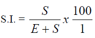
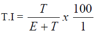
where S = numbers of stomata and E = epidermal cells within the particular area under investigation. The same
formula was applicable for the calculation of Trichome Index (T.I.), in this case, trichome (T) was used instead of
stomata:
Anatomical Studies: Seeds of the plant materials were plated out in petri dishes containing moist Whatman
(110mm) filter paper and the germination test was calculated using similar formula as applied to stomatal index but
based on the percentage of the number that germinated divided by total number of seeds plated. Some seeds were
planted in labelled containers. Three days to two weeks after growth had occurred, stem and root systems were
fixed, alongside with mature leaves, flowers, fruits and petioles harvested from mature plant, in FAA in the ratio of
1:1:18 of 40% formaldehyde, glacial acetic acid and 70% alcohol for at least 48% hours following the method of
Johansen [11].
Free hand sectioning using a systematic arrangement of 5 razor blades, with 2 sets ( nacet and tiger blades) crossed
and a central vertical one (nacet) lying in between the 2 sets crossed. The blades were adjusted until the holes in
them synchronized. The plant part to be sectioned was placed in the hole and using the first two fingers of the left
hand to hold the vertical blade sets, while pressing down the 2 crossed sets with the first two fingers of the right
hand to make a transverse section of about 20 to 25μm thick. The sections made were passed through alcohol
solutions in the order: 30%, 50% 70%, 95% and absolute alcohol, allowing them for 5 minutes in each solution. The
dehydrated materials were cleared of their natural wax by passing them through different proportions of alcohol and
chloroform in the following ratios (3:1; 1:1; 1:3) v/v for 10 minutes in each, and as the chloroform gradually
replaced the alcohol, the process was repeated from the pure chloroform and down the series again within same time
interval. These were rehydrated in alcohol series starting with absolute then 95%, 70%, 50%, 30% and stained with
1% Alcian blue for 2 minutes, washed off with water before counter-staining with 1% safranin for 2 minutes. The
stain was washed off and placed on clean glass slide with a drop of glycerol and a clean cover slip placed on it. The
slides so prepared are as good as those of microtomy and are near permanent ones. These slides were viewed with
the light microscope and photomicrographs taken from good preparations after proper examination.
Cytological Study: Healthy root tips for mitotic study were obtained from seeds of Capsicum annuum Linn. grown
in a petri dish containing 110mm Whatman filter paper moist with water for a period of three days to one week. The
early germinated roots were transferred to solution of 0.002M of 8- hydroxyquinoline for 3 hours specifically to
suspend the spindle fibres or to accumulate chromosomes at metaphase between 9 and 10 a.m. to be precise. The
roots were treated with Carnoy’s fluid (3:1 ethanol/acetic acid v/v) for 12 to 24 hours aimed at killing the cells. The
roots were then preserved in 70% alcohol and kept in the refrigerator until when needed or used immediately by
hydrolyzing in 9% HCl for 8 minutes and passing them through 70% ethanol for 10 minutes. 1mm of the root tip
studied was excised from the apex and squashed in a drop of FLP-orcein stain( 2g of orcein dissolved in 100ml of a
solution of equal parts of formic acid, lactic acid, propanoic acid and water) under a cover slip, flattened out and
examined under a light microscope, following the method of Okoli [12]. Photomicrographs of the chromosomes
were taken from good temporary slides, using a Sony digital camera (7.2 Mega pixels).
Phytochemical Studies (Qualitative analyses): The leaves of the species were sun dried for 72 hours (3 days) and
weighed. Fifty grams (50g) of the leaves were macerated in 96% ethanol using a pestle and a mortar. The extract
was there after filtered and evaporated to dryness using a rotary evaporator set at 45°C to constant weight and later,
an exhort extraction machine. Residue yields were noted and a portion was used for the phytochemical screening.
Phytochemical screening for saponin, frothing tests, was done following the method described by Wall et al. [13,14] as shown below:
The ability of saponins to produce frothing in aqueous solution and to haemolyse red blood cells was used as
screening test for these compounds. 0.5g of each plant extract was shaken with water in a test tube. Frothing which
persisted on warming was taken as preliminary evidence for the presence of saponins. In order to remove ‘falsepositive’
results, the blood haemolysis test was performed on those extracts that frothed in water. 0.5g of each
extract was boiled briefly with 50ml phosphate buffer, pH 7.4, and then allowed to cool and filtered; 5ml of the
filtrate was passed for 3 hours through an asbestos disc (1.5mm thick and about 7mm in diameter), which had been
previously soaked with two or three drops of 1 percent cholesterol in ether and dried. After filtration the disc was
washed with 0.5ml of distilled water, dried and boiled in 20ml of oxylol for 2 hours to decompose the complex
formed between cholesterol and any saponins in the extract. The disc was then washed in ether, dried and placed on
a 7 percent blood nutrient agar. Complete haemolysis of red blood cells around the disc after 6 hours was taken as
further evidence of presence of saponins.
Test for alkaloids: 0.5g of each extract was stirred with 5ml of 1 percent aqueous hydrochloric acid on a steam bath;
1ml of the filtrate was treated with a few drops of Mayer’s reagent and a second 1ml portion was treated similarly
with Dragendorff’s reagent. Turbidity or precipitation with either of these reagents was taken as preliminary
evidence for the presence of alkaloids in the extract being evaluated [15, 16]. A confirmatory test designed to
remove non-alkaloidal compounds capable of eliciting ‘false-positive’ results was carried out as follows with all
extracts which gave preliminary positive tests for alkaloids. A modified form of the tin-layer chromatography (TLC)
method as described by Farnsworth and Euer [17] was used. 1g of the extract was treated with 40 percent calcium
hydroxide solution until the extract was distinctly alkaline to litmus paper, and then extracted twice with 10ml
portions of chloroform. The extracts were combined and concentrated in vacuole to 5ml. The chloroform extract was
then spotted on thin-layer plates. Four different solvent systems (of widely varying polarity) were used to develop
each plant extract. The presence of alkaloids in the developed chromatograms was detected by spraying the
chromatograms with freshly prepared Dragendorff’s spray reagent. A positive reaction on the chromatograms
(indicated by an orange or darker coloured spot against a pale yellow background) was confirmatory evidence that
the plant extract contained an alkaloid.
Test for tannins: 5g of each portion of plant extract was stirred with 10ml of distilled water, filtered, and ferric
chloride reagent added to the filtrate. A blue-black, green, or blue-green precipitate was taken as evidence for the
presence of tannins [16].
Test for anthraquinones: Borntrager’s test was used for the detection of anthraquinones. 5g of each plant extract was
shaken with 10ml benzene, filtered and 5ml of 10 per cent ammonia solution added to the filtrate. The mixture was
shaken and the presence of a pink, red, or violet colour in the ammonia (lower) phase indicated the presence of free
hydroxyanthraquinones.
For combined anthraquinones, 5g of each plant extract was boiled with 10ml aqueous tetraoxosulphate (vi) acid and
filtered while hot. The filtrate was shaken with 5ml of benzene, the benzene layer separated and half its own volume
of 10 per cent ammonia solution added. A pink, red, or violet coloration in the ammonia phase (lower layer)
indicated the presence of anthraquinone derivatives in the extract [16].
Test for phlobatannins: Deposition of a red precipitate when an aqueous extract of the plant part was boiled with 1
per cent aqueous hydrochloric acid was taken as evidence for the presence of phlobatannins [16].
Test for cardiac glycosides: Lieberman’s test was used. 0.5g of the extract was dissolved in 2ml of acetic anhydride
and cooled well in ice. Tetraoxosulphate (vi) acid was carefully added. A colour change from violet to blue to green
indicated the presence of a steroidal nucleus (i.e. aglycone portion of the cardiac glycoside) [18].
Results
Macro-morphology
The geographic location of the parent plant studied was 040521337711N and 006054186011E at 18m altitude. The
opening and closing times of the flowers were studied. It was revealed that the flowers commence opening at 6:50
a.m. and open completely at 8:30 a.m. while the closing time start at 5:00 p.m. and close completely at 8:50 p.m.
.These features are of taxonomic relevance. The germination test conducted was 60%. The distributional pattern of
the species has been recorded by Hutchinson and Dalziel [3]. Pepper as commonly known is an annual stout
branched sub woody plant attaining up to a height of 65cm. The sub-sessile leaves are simply ovate, apex acutely
acuminate and cuneate or abruptly acute at base measuring up to 19cm in length and 4cm wide. The petioles are
0.2cm in length. The glabrous stem is also angular in shape. Inflorescence is terminal and flowers are borne singly at
nodes and measuring up to 0.4cm in diameter. The petals are 5 and 6 in number, pale whitish and averagely 0.5cm
long and 0.3cm wide. The greenish sepals are 5 and 6 in number but not separated, averagely 0.2cm long and 0.1cm
wide. The stamens are also 5 and 6 in number up to 0.3cm long. It is discovered that the plant is both pentamerous
and hexamerous. The fruits comprised many seeded berry, borne singly at nodes, globose in shape and when unripe
green, and when ripe are red, orange, yellow, brown, or purplish up to 1 to 2.5cm in diameter. The seeds are 0.2 to
0.3cm in diameter (Figure 1). The morphology and quantitative characteristic of the plant is revealed Tables 1 and 2.
Aestivation type for the species studied is valvate. Insect pollinators are ants, spiders, house flies, bees and
caterpillars. Pollinators started appearing at 7:00 a.m. and were not seen at 2:20 p.m., and sometimes resurfaced later
in the day.
Figure 1: Capsicm. Annuum Linn. Arrow shows hexamerous flower
TABLE-1: Morphological Characteristics of Capsicum annuum Linn.
TABLE-2: Quantitative Characteristics of Capsicum annuum Linn.
Micro- morphology
Capsicum annuum Linn. foliar epidermal study revealed the presence of anomocytic stomata and uniseriate
trichomes at both the adaxial and abaxial foliar surfaces Tables 3 and 4. It is shown that the adaxial foliar layer has
6.02% stomatal index and 25.95% for the abaxial surface, in other words, there are more stomata in the abaxial
foliar surface than as observed in the adaxial foliar surface, see Figures 2 and 3. Trichome index is also studied
revealing 1.61% for the adaxial and 7.69% for the abaxial surfaces. Stem epidermal study showed paracytic stomata,
uniseriate trichomes and irregularly-shaped cells (Figure 4). The stomatal characteristics showed that the adaxial
stomatal length of 12.7±3.20μm with 25.18% coefficient of variation (C.V.) and width of 8.70±1.98μm with 21.71%
C.V., while the abaxial stomatal length of 12.0±1.83μm with 28.56% C.V. and width of 7.6±1.27μm with 16.64%
C.V. (Table 5).
TABLE - 3: Stomatal Indices of Capsicum annuum Linn.
TABLE - 4: Trichome Indices of Capsicum annuum Linn.
Figure 2: Adaxial surface Arrow reveals glandular trichome
Figure 3: Abaxial surface- Anomocytic stomata
Figure 4: Stem epidermis- Arrow shows biseriate trichome
TABLE-5: Stomatal Characteristics of Capsicum annuum Linn.
Anatomy
Table 6 gives the summary anatomy of Capsicum annuum Linn. The mid-rib shows uniseriate trichomes in
epidermis made of a layer of cells. The collenchymatous cells occupy the region of the hypodermis. Parenchymatous cells occupy the ground meristem. The primary growth phase reveals 2 vascular traces. The vascular bundles have
bicollateral arrangement, no rib bundle wings observed in the mid-ribs of both primary and secondary growth phases
(Figure 5). The petiole of Capsicum annuum Linn. is made of a layer of cells in the epidermis, 2 to 4 layers of
collenchyma in the hypodermis, the general cortex is predominated by parenchymatous cells. The primary growth
phase revealed 2 vascular traces and 2 rib bundle wings observed at each wing position (Figure 6). Internodal anatomy
of Capsicum annuum Linn. showed a four-sided or rectangular structure with swollen protuberances at each end.
The hypodermis is made of 5 layers of collenchymatous cells, and the general cortex has 3 layers of parenchyma of
thin walls. The endodermis is made of a layer of barrel-shaped cells. The pericycle just below the endodermis is
composed of 3 to 4 cell-layers. The pith region is made of large parenchymatous cells (Figure 7). The nodal pattern is
unilacunar having 2 vascular traces from 1 gap (Figure 8). Root anatomy of Capsicum annuum Linn. revealed the
piliferous layer as single-cell thick. The vascular bundles are radially symmetrical with exarch xylary cells.
Centralized parenchymatous cells occupy the pith region of the root (Figure 9). Ovary anatomy of Capsicum annuum Linn. revealed the placentation as axile type . It is bilocular and 2-celled (Figure 10).
TABLE- 6: Anatomy of Capsicum annuum Linn.
Figure 5: Mid – rib anatomy pattern unilacunar. Double – way arrow between 2 vascular traces
Figure 6: Petiole anatomy. Arrow point at vascular arc
Figure 7: Stem inter modal anatomy
Figure 8: Nodal; arrow reveals 2 vascular traces from 1 gap
Figure 9: Root anatomy with vascular system at radial symmetry
Figure 10: Ovary anatomy; arrow at the position of placenta
Cytology
Cytological Studies of Capsicum annuum Linn. showed the mitotic chromosome number as 2n=24 at side
metaphase (Figure 11). The karyotype revealed 2 telocentric chromosomes (Figure 12).
Figure 11: Mitotic chromosomes. Arrow reveals a set of Telecentric chromosome
The phytochemical studies revealed the presence of the following secondary metabolites: alkaloids, saponins,
tannins, phlobatannins, flavonoids, combined anthraquinones, free anthraquinones and cardiac glycosides
respectively (Table 7).
TABLE-7: Phytochemical Properties of Capsicum annuum Linn.
Discussion
Observations on vegetative and floral features of Capsicum annuum Linn. reveal the habit of the species as either
annual herbs or short-lived perennial sub-woody plant which is of taxonomic relevance. Capsicum annuum Linn. is
mostly annual sub-shrub as also recorded by Hutchinson and Dalziel [3]. Fruits of Capsicum annuum Linn. are very
pungent and as such are used as spices and preservatives, as supported by Gill [2]. Capsicum annuum Linn.
possesses simple uniseriate trichomes. The stem epidermal study revealed paracytic stomata. Capsicum annuum Linn. was observed having pentamerous and hexamerous flowers. The structure of the stamens and carpels, and mostly their pilose nature are of taxonomic importance in the delimitations at generic and species level. The stem
investigation revealed Capsicumannuum Linn. as angular or tetra-angular structure, also possessing swollen
protuberances at the tetra angular ends. The primary node of Capsicum annuum Linn. Is unilacunar and the roots
have radially symmetrical vascular system. The species investigated is bisexual, hypogynous and placentation is
axile which is also in accordance to the observation of Hutchinson and Dalziel [3]. The fruit is globose as supported
by the findings of Hutchinson and Dalziel [3].
Anatomically, studies on the primary growth phase revealed the mid-rib and petiole of Capsicum annuum Linn. are
observed with 2 vascular traces and also have bicollateral vascular system. It was observed that the departure of the
rib-bundle wings is towards the position of the open vascular system. The secondary growth phase revealed vascular
arc structure in the mid-rib and petiole, while the stem and root showed a complete ring structure of an open
vascular system.
Cytologically, the basic chromosome number for members of Solanaceae is x = 12 Omidiji [7] Okoli and Osuji [8]
also supported the chromosome basic number as x = 12, and diploids of 2n = 24.The presence of phytochemical
constituents account for the therapeutic effect of the plant in trado-medicine.
Acknowledgement
I appreciate my project supervisor Prof. B. E. Okoli for his guidance and advice. My honest gratitude to Prof. (Mrs)
L. A. Akonye for her indelible role in my career and DR. N.L. Edwin-Wosu for the authentication of the plant
species in the Herbarium. My unreserved thanks to my wife for her encouragement and support. Above all things, I
thank my Lord and Saviour Jesus Christ, whose wonderful protection, love and grace have strengthened me at all
times.
References
- Watson, L. and Dallwitz, M.J. The Families of Flowering Plants. Descriptions illustrations, identification and information. Retrieval version ALPHA35 <https://deltantkey.com/angio/www/Solanaceae.htm.> (Accessed 9August, 2012) 1992.
- Gill, L.S. Taxonomy of flowering Plants Africana-Fep. Publishers Ltd., Nigeria. 1987.
- Hutchinson, J. and Dalzial, J.M. Flora of West Tropical Africa .Vol, 1, Part 2, Crown agents for Overseas Governments and Administration London. 1958.
- Purseglove, J.W. Tropical cropsdictyledons. Longman Inc.New York. 1968.
- Metcalfe, C.R. and L. Chalk. Anatomy of the Dicotyledons. 2nd eds. Oxford: The Clarendon Press. 1979.
- Cutler, D. F. Applied Plant Anatomy. Longman-Group Limited, London. 1977.
- Omidiji, M.O, Nigerian Journal of Genetics, 1985, 6:75- 83.
- Okoli, B. E. and Osuji J.O. Nigerian Journal of Botany, 2008, 21(2):358-372.
- Cutler, D. F. Applied Plant Anatomy.Lib.of Congr.Cataloguing in Publication Data. William Clowes and Sons Ltd. London. 1978.
- Arnold, E. Peacock’s Elementary Microtechnique. Pitman Press, Bath, Great Britain. 1973.
- Johansen H. Plants Microtechnique. McGraw-Hill, New York. 1940.
- Okoli, B.E. New phytol. 1983, 93: 59 – 97.
- Wall, M. E., Eddy, C. R., McClenna, M. L. and Klump, M. E. Anal Chem. 1952. 24: 1337.
- Wall, M. E.,Krider, M. M., Krewson, C. F., Wilaman, J. J., Cordell, D. S. and Gentry, H. S.’Steroidal sapgenins X111. Supplementary table of data for steroidal sapogenins V11’. 363 pp. 1954.
- Harborne, J. B, Phytochemical Methods: A Guide to Modern Techniques of Plant Analysis. Chapman & Hall,London.279 pp 1973.
- Trease, G. E, and Evans, W. C. A Text Book of Pharmacognosy. 3rd eds. Boilliere Tinall Ltd., London. 1989.
- Farnsworth, N. R. and Euer, K. LAn Lioydia.25-186. .1962.
- Shoppee, C. W. Chemistry of the Steroids, 2nd edn.Butterworths, London. 1964.


