Keywords
Progesterone, Vitamin E, Pregnancy, Embryo, Progesterone profile, Young rabbit
Introduction
Rabbit are widely used for human consumption and in reproductive medical experiments. After fertilization, P4 produced from the corpus luteum is known to prepare the uterine lining for zygote implantation and to prevent the uterine contractions for maintenance of pregnancy. Embryo survival is dependent on the uterine environment and composition of its fluid during the luteal phase prior to implantation. Thus, a relationship between P4 concentration released in the early luteal phase and embryo loss [1] has a physiological basis in uterine growth and suppression of myometrial contractility [2]. Also, a delay in P4 secretion during the early post-ovulatory luteal phase had a negative impact on embryo survival [3]. Similarly, daily injections of 25 mg P4 from 36 h post-mating in ewes increased blastocyst diameter by 220% on day 9 and advanced the time of elongation of blastocysts to a filamentous conceptus [4,5]. However, young does that gestate at first time are usually poor in P4 production due to inadequate CL development.
Antioxidant vitamins, alone or combined with other supplements, decrease embryonic mortality and improve birth outcomes [6]. Previous studies [7,8] indicated that oxidative damage on DNA, protein, and lipids during pregnancy may be associated with reduced birth weight and increased risks of preterm delivery and preeclampsia. Hence, filling reproductive tissues of pregnant dames by potential antioxidants can protect the maternal-fetal unit and increase the intrauterine growth and infant weight at birth. Vitamin E is a lipid soluble, chain-breaking antioxidant that is dietary in origin. Vitamin E has potential consequence on the outcome of pregnancy via prostacyclin [9], where vitamin E enhanced the release of prostacyclin, it metabolite of arachadonic acid, which inhibits platelet aggregation, retarded uterine contractility, and increased vasodilation [9]. Both α- and γ-tocopherols could be involved in the fetal growth by increasing blood flow to the fetus. Therefore, the objective of this study is to investigate the effect of P4 and Vitamin E injections and their integration on pregnancy enhancement, embryo development and peripheral P4 concentrations in young rabbit does.
Materials and Methods
The experiment was carried out in the Animal House, Faculty of Medicine, Assiut University, Assiut, Egypt in early spring.
Animals
A total of 24 virgin female WNZ rabbit does (6 months of age) with a mean body weight of 2.35 ± 0.49 kg were divided randomly into four groups of six does per each. Rabbits were vaccinated to protect animals from bacterial septicemia and viral hemorrhagic diseases. Does in the first group were injected i.m. with saline solution 2 days after natural mating and considered as the control for the treated groups. Does in the second group were injected i.m. with a single dose of 1.0 mg long-acting progesterone (P4, hydroxyl-progesterone caprote, Cidolut Depot, CID, Egypt) 2 days after natural mating. Does in the third group were injected i.m. with 20 IU Vitamin E (Pharco Pharmaceutical, Alex, Eg.) per kg body weight every 3 days after natural mating until mid-pregnancy (day 15). Does in the fourth group were injected i.m with both P4 and Vitamin E.
Feeding
All does were fed a diet containing 18.25% crude protein, 2.29% crude fat (2700 kcal/kg ration metabolizable energy (ME) according to [10] and 13.4% crude fiber. The amount of Vitamin E in this diet was 30-50 mg/kg diet. Ingredient constituents of the diet were yellow corn, wheat bran, soybean meal (44%), dicalcium phosphate, limestone, sodium chloride, methionine, premix, molasses and alfalfa hay. Green roughage and water were supplied ad libitum throughout the experiment. Each doe was fed ad libitum on this commercial diet throughout the experiment.
Housing
Rabbits were reared in individual cages (50 × 60 cm × 35 cm) of galvanized wire net, equipped with an automatic drinker and a manual feeder. These cages were put in rooms with comfortable environmental conditions, the temperature and humidity inside the room (31 m × 8 m × 3 m) were 19°C-22°C and 75%-80%, respectively. Temperature and humidity were maintained using an air conditioner-heater system and forced ventilation to remove gases such as carbon dioxide. Does were housed under 14 h light: 10 h dark throughout the experiment.
Mating
All does were mated naturally by WNZ fertile bucks selected on basis of vigor, physical conformation and semen quality. The rabbit doe was brought to the buck’s hutch, mated during a 10 min interval and re-mated within 4 h after the initial mating. This short interval between mating may increase the probability of conception as remating 36 h after the initial mating may cause undesirable alteration of estrogen and P4 in this induced ovulatory and lead to abortion or miscarriage.
Blood Collection
Blood samples were withdrawn from all does by using 25-gauge needle attached with 3 cc syringes during premating, 7 days post-mating and mid-pregnancy (total of 72 samples). Approximately 3 ml blood was collected from each doe via the central ear vein. Before collection, one drop of Xylol was spread on the ear by using small clean cotton for increasing the blood flow in the ear veins [11]. Blood samples were allowed to clot at 4°C overnight in the refrigerator and centrifuged at 2000 × g for 20 min. After centrifugation, sera were collected in 4 ml eppendorf tubes and stored at -20°C until P4 assay.
Progesterone (P4) Assay
P4 was assayed by competitive immunoluminometric assay (Maglumi Prog, Shenzhen New Industries Biomedical Engineering Co. Ltd, London, UK). This method was based on the principle of competitive binding with an assay range of 0-80 ng/ml.
Ultrasonographic Examination
Ultrasonography was used in this experiment for pregnancy diagnosis and determination of embryo development on days 7, 10, 12, 14, 16, 18, 20 and 22 after mating. After shaving hair from the belly, the uterus of each doe was examined trans-abdominally by Doppler ultrasonographic technique (ESAOTE/Pie Medical My Lab 30 vet deviceviadi Caciolle,15-50/27, Frienze, Italy) [12]. This method was equipped on 6 MH2 and the probe was positioned externally against the abdominal wall while the doe was restrained in a dorsal recumbent position. Diagnosis of pregnancy was confirmed by palpation on day 12 after mating according to [13].
Statistical Analysis
Data of were analyzed statistically (one and two way analysis of variance) using the GLM procedure of SPSS software [14]. 20 Differences between LSM were analyzed by using [15]. Data in percentage values were transformed with the arcsine square root procedure to normalize the variance before analyzing the data.
Results
Embryo and Fetus Development
On day 7 after mating, there were no significant differences in uterine diameters of non-pregnant treated groups and controls one. The non-gravid uterus contained hypoechogenic (dark grey) linear structure with diameter 0.34 ± 0.09 cm without any contents in the lumen (Figure 1), where the percentages were 11%, 50% and 25% in P4, Vitamin E and P4+Vitamin E) Compared with the control one (45%). While In pregnant does, the gravid uterus of pregnancy contained an echogenic (black) amniotic vesicle in the uterine lumen with hyperechogenic (white) eccentric masses which represents the fetuses (Figure 2). Pregnancy rates were 89%, 50% and 75% in P4, Vitamin E and P4+Vitamin E compared with 55% in the control. There were significant differences between the P4 group and control group (P<0.05). The amniotic vesicle diameter was larger in P4 group of pregnant does (0.89 ± 0.34 cm) compared with the control (0.53 ± 0.11 cm). Otherwise, amniotic vesicle diameter was not affected by treatment of Vitamin E alone (P>0.05). Doppler analyses did not detect significant differences in the blood supply perfusion due to treatments and the controls.
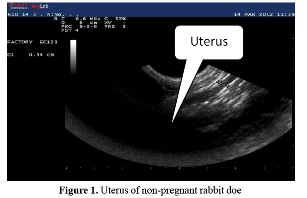
Figure 1. Uterus of non-pregnant rabbit doe
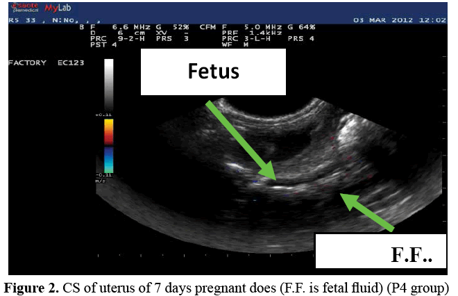
Figure 2. CS of uterus of 7 days pregnant does (F.F. is fetal fluid) (P4 group)
On day 10, the gravid uterus contained anechogenic (black) amniotic vesicles in the lumen with larger hyperechogenic mass than observed on day 7 of pregnancy. The number of fetuses could be counted (Figure 3) (P<0.05) in P4 and control groups. Amniotic vesicles diameters were again larger in P4 group (1.19 ± 0.11 cm) compared with the control (0.71 ± 0.09 cm; P<0.05). Treatment with vitamin E again did not affect amniotic vesicle diameter.
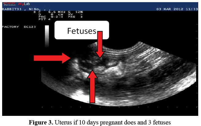
Figure 3. Uterus if 10 days pregnant does and 3 fetuses
On day 12, the diameter of the amniotic vesicle was larger than that observed on day 10 after mating. Moreover, blood supply of fetus and uterus increased in this period (Figure 4). Also, diameters of amniotic vesicles contained fetuses were significantly (P<0.05) larger in P4 and P4+Vitamin E groups than Vitamin E group and control one (2.31 ± 0.43 cm vs. 1.63 ± 0.28 cm, respectively) where, Vitamin E group was similar to the control one.
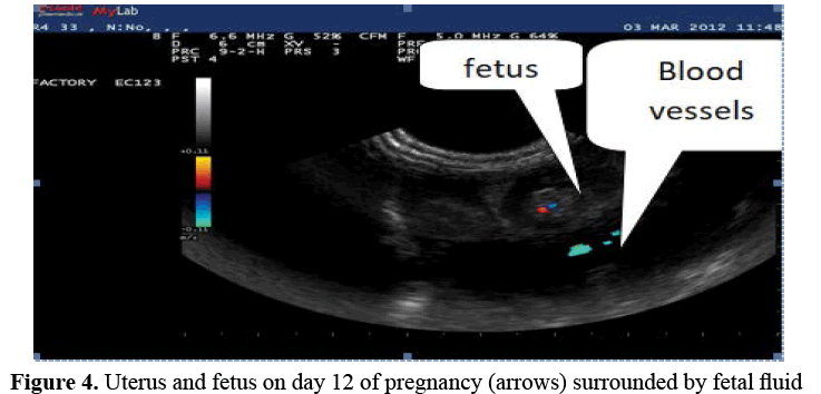
Figure 4. Uterus and fetus on day 12 of pregnancy (arrows) surrounded by fetal fluid
On day 14, the fetus was more visible in the uterus with heart beats detectable in all fetuses, where, crown rump length (fetus length) was 1.89 ± 0.36 cm, 1.78 ± 0.22 cm, and 1.30 ± 0.17 cm in P4, Vitamin E+P4 and Vitamin E groups vs. 1.23 ± 0.39 cm in the control. This fetal size was significantly (P<0.05) larger in P4 and P4+Vitamin E groups than in the Vitamin E group and the control one. Blood supply was not significantly different among the studied groups. The number of fetuses could be counted in that period.
On day 16, fetus length continued to be larger (P<0.05) in both P4 and Vitamin E+P4 groups (1.94 ± 0.25 cm and 1.86 ± 0.36 cm) compared with the Vitamin E group and the control (1.53 ± 0.11 cm and 1.44 ± 0.27 cm. Number of fetuses could be counted during that period, but there were no significant differences in the blood supply among the groups.
On day 18, fetuses occupied all spaces of uterus; hence, therefore, number of fetuses could not be counted easily. Fetus length was significantly longer in P4 group (2.54 ± 0.33 cm) than in other groups with averaged lengths of 2.01 ± 0.16 cm in Vitamin E+P4, 1.94 ± 0.23 cm in Vitamin E and 1.89 ± 0.14 cm in the control (Figure 5). Blood supply was not affected by treatments.
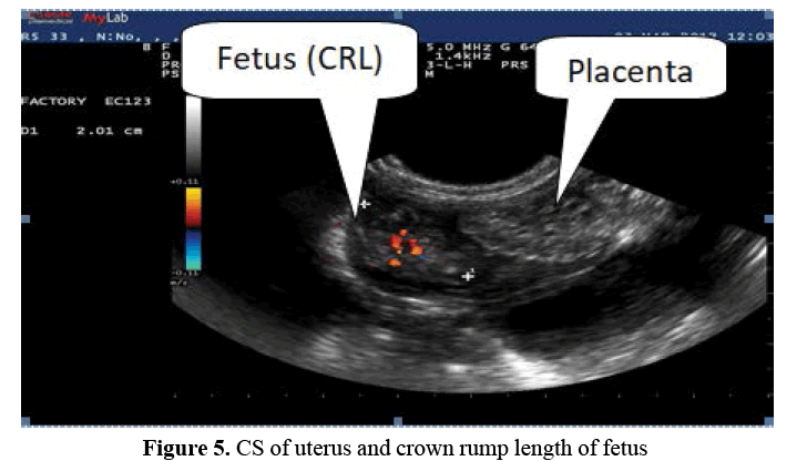
Figure 5. CS of uterus and crown rump length of fetus
On day 20, fetuses could not be counted, but fetus length increased significantly in P4 group (2.83 ± 0.21 cm) compared with the other groups 2.22 ± 0.31 cm (Vitamin E+P4), 2.08 ± 0.27 cm (Vitamin E) and 1.99 ± 0.14 cm (control) (Figure 6). Fetal parts such as trunk and head could be distinguished (Figure 7). While, blood supply was not affected by treatments.
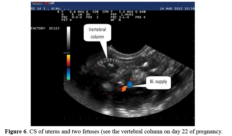
Figure 6. CS of uterus and two fetuses (see the vertebral column on day 22 of pregnancy.
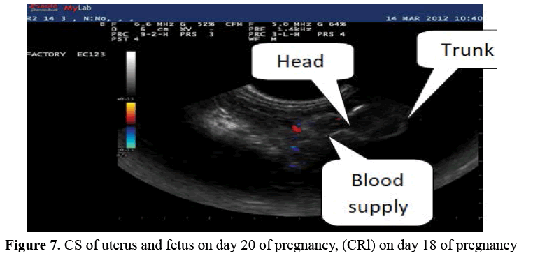
Figure 7. CS of uterus and fetus on day 20 of pregnancy, (CRl) on day 18 of pregnancy
Similarly, on days 20 and 22, fetuses occupied the whole uterine lumen with relatively small amounts of fetal fluid surrounding the fetuses. Fetus length was larger in P4 group (3.64± 0.53 cm) compared with the other groups 3.03 ± 0.49 cm (Vitamin E+P4), 3.07 ± 0.36 cm (Vitamin E) and 2.98 ± 0.33 cm controls (Figure 8). Fetal parts such as vertebral column and ribs could be distinguished in Figure 7, while blood supply was not influenced by treatments.
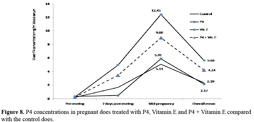
Figure 8. P4 concentrations in pregnant does treated with P4, Vitamin E and P4 + Vitamin E compared with the control does.
P4 Concentration
P4 concentration was significantly higher at one week of pregnancy in P4 group (4.97 ± 1.27 ng/ml blood serum) than the other treatments. P4 concentration continued to rise to reach 12.45 ± 2.79 ng/ml in mid-gestation of does treated by P4 compared with control and Vitamin E groups. Moreover, P4 concentration did not differ between the does treated with Vitamin E and the controls Figure 8. While, P4 concentration in combined group was similar to that in P4 group.
Discussion
Synchrony between mammalian embryo implantation and uterine development are essential for subsequent development of embryos. Rabbit embryos can tolerate up to two days of asynchrony [16,17]. Administration of P4 can advance the secretion of characteristic uterine proteins in the rabbit [18]. Our results revealed that P4 alone or in combination with Vitamin E facilitated establishment of pregnancy and maintained embryonic survival during early stage of pregnancy (first week) these results are coincident with findings obtained by [3,19] who indicated that P4 concentration might be related to the embryo survival and low P4 concentrations during the early post-ovulatory luteal phase or a delay in its normal rise in that period were associated with reducing embryo survival. Further, lack of corpora lutea reduced embryo survival in rabbits [19]. This means that use of P4 in the current study could protect embryos from mortality presumably either by reducing myometrial contraction or by maintaining maternal extracellular fluid volume during pregnancy, where [20] indicated that rate of oviduct fluid secretion was regulated by ovarian hormones and uterine milieu during early pregnancy contained at least a 3-fold increase in nutrients compared with non-pregnant animals [21]. Antioxidant vitamins alone or combined with other supplements, decreased embryonic mortality and improved birth outcomes [6,22]. There are contradictory results in regard to the role if Vitamin E on fetal growth [23,24]. However, the increased amniotic vesicle diameter observed in the present study may due to effect of P4 dose; this is similar to the increased growth of day 9-12 embryos stimulated by P4 supplementation in sheep [4]. Also, P4 administration led to increased fetal length on days 14, 16 and 22, this may be attributed to effects of P4-induced changes in gene expression of uterine tissues [4,25,26] and changes in composition of histotroph surrounding developing embryo. In cows, administration of P4 at days 1-4 of pregnancy enhanced fetal development (measured on day 14) compared with control [27,28]. Similarly, ewes received P4 on day 1-3, 3-6 or 1-6 of pregnancy had higher plasma P4 than the control and fetal growth was greatest when the treatment started on day 1 of pregnancy [29]. Previous study [30] reported that embryo survival was associated with large variations in P4 secretion during first few days after ovulation and the high P4 concentrations were related to the embryo survival. In the present study, P4 concentrations in does treated with P4 and P4+Vitamin E was higher than that treated with Vitamin E only and the controls. Previous study [31,32] reported that the increased P4 concentrations on days 12 and 24 of pregnant rabbits may be attributed to the positive impact of Vitamin E and Se intake on P4 concentration. Further, P4 concentrations increased in the second half of pregnancy on days 12 and 13 with a peak on days 14-17 [32].
Conclusion
P4 alone or combined with Vitamin E maintained embryo survival and pregnancy in young does. Vitamin E alone did not have a significant effect. Hence P4 and P4+Vitamin E may be applied successfully to enhance pregnancy in rabbit does and in utero development of offspring.
Acknowledgement
Authors would like to thank Assiut University for funding this research article.
References
- Morris D, Diskin M. Effect of progesterone on embryo survival. Animal. 2008;2(8):1112-1119.
- Graham JD, Clarke CL. Physiological action of progesterone in target tissues. Endocr Rev. 1997;18(4): 502-519.
- Diskin MG, Morris DG. Embryonic and early foetal losses in cattle and other ruminants. Reprod Domest Anim. 2008;43:260-267.
- Satterfield MC, Bazer FW, Spencer TE. Progesterone regulation of preimplantation conceptus growth and galectin 15 (LGALS15) in the ovine uterus. Biol Reprod. 2006;75(2):289-296.
- Lamming GE, Royal MD. Ovarian hormone patterns and subfertility in dairy cows. Proceedings of an International Symposium Organized by the British Society of Animal Science Entitled ’Fertility in the High- Yielding Dairy Cow’, Galway, Ireland, September 1999. BSAS, Occasional Publication 2001;26(1):105-118.
- Cederberg J, Siman M, Eriksson UJ. Combined treatment with vitamin E and vitamin C decreases oxidative stress and improves fetal outcome in experimental diabetic pregnancy. Pediatric Res. 2001;49(6):755-762.
- Chappell LC, Seed PT, Briley A, et al. Effect of antioxidants on the occurrence of preeclampsia in women at increased risk: A randomized trial. Lancet. 1999;354(9181):810-816.
- Scholl TO, Leskiw M, Chen X, et al. Oxidative stress, diet and the etiology of preeclampsia. Am J Clin Nutr. 2005;81(6):1390-1396.
- FNB. Food and Nutrition Board, Institute of Medicine. Dietary reference intakes for vitamin C, vitamin E, selenium and carotenoids. Washington, DC: National Academy Press, 2000.
- NRC. National Research Council. Nutrition requirements of domestic rabbits. National Academy of Sciences 1977: Washington DC, USA.
- Hoppe PC, Laird CW, Fox RR. A simple technique for bleeding the rabbit ear vein. Lab Anim Care. 1969;19(4):524-525.
- Gutierrez HE, Zamora FMM. Ultrasonography study of rabbit’s pregnancy. Proceedings - 8th World Rabbit Congress -September 7-10, 2004 - Puebla, Mexico.
- Ypsilantis P, Saratsis PH. Early pregnancy diagnosis in the rabbit by real time ultrasonography. World Rabbit Sci. 1999;7(2):95-99.
- SPSS. Statistical package for social sciences. Release 17.0 SPSS Inc. 2008; USA.
- Duncan DB. Multiple range and Multiple F-tests. Biometrics. 1955;11(1):1-42.
- Adam CE. Ovarian control of early embryonic development within the uterus. In: Lamning, G.E. and Amoroso, EC, edition. Reproduction in the Female Mammal Buttemorth, Iondon; 1968;532-546.
- Chang HC. Development and fate of transferred rabbit ova or blastocysts in relation to the ovulation time of the recipients. J Exp Zool. 1950;114(1):197-216.
- McCarthy SM, Foote RH, Mmrer RR. Embryo mortality and altered uterine luminal proteins in progesterone-treated rabbits. Fert and Steril. 1973;28(1):101-111.
- Battista MG, Pope WF, Foote RH. Plasma and uterine progesterone and embryo survival in rabbits following asynchronous transfer to unilaterally ovariectomized recipients. Theriogenology. 1987;27(6):897-905.
- Black DL, Duby RT, Riesen J. Apparatus for continuous collection of sheep oviduct fluid. J Reprod Fertil. 1963;6:257.
- Gao H, Wu G, Spencer TE, et al. Select nutrients in the ovine uterine lumen. I. Amino acids, glucose, and ions in uterine lumenal flushings of cyclic and pregnant ewes. Biol Reprod. 2009;80(1):86-93.
- Coffey MT, Britt JH. Enhancement of sow reproductive performance by beta-carotene or vitamin A. J Anim Sci. 1993;71(5):1198-202.
- Valsecchi L, Cairone R, Castiglioni MT, et al. Serum levels of alpha-tocopherol in hypertensive pregnancies. Hypertens Pregnancy. 1999;18(3):189 -195.
- Tamura T, Goldenberg RL, Johnston KE, et al. Serum concentrations of zinc, folate, vitamins A and E, and proteins, and their relationships to pregnancy outcome. Acta Obstet Gynecol Scand. 1997;76: 63-70.
- Bauersachs S, Ulbrich SE, Gross K, et al. Embryo-induced transcriptome changes in bovine endometrium reveal species-specific and commonmolecular markers of uterine receptivity. Reprod. 2006;132(2):319-331.
- Forde N, Carter F, Fair T, et al. Progesterone-regulated changes in endometrial gene expression contributes to advanced conceptus development in cattle. Biol Reprod. 2009;81(4):784-794.
- Machlin LJ. Vitamin E in handbook of vitamins: Nutritional, biochemical and clinical aspects. Marcel Dekker, Ney York, Inc. 1984;117(2):99-145.
- Garrett JE, Geisert RD, Zavy MT, et al. Evidence for maternal regulation of early conceptus growth and development in beef cattle. J Reprod Fertil. 1988;84(2):437-446.
- Kleemann DO, Walker SK, Seamark RF. Enhanced fetal growth in sheep administered progesterone during the first three days of pregnancy. J Reprod Fertil. 1994;102(2):411-417.
- Ashworth CJ, Sales DI, Wilmut I. Evidence of an association between the survival of embryos and the periovulatory plasma progesterone concentration in the ewe. J Reprod Fertil. 1989;87(1):23-32.
- Abou-Zeina HAA, Hamam AM. Effect of parental administration of vitamin E and or Selenium on health and reproductive function of ewes fed on marginal deficient diets. Egypt J Basic Appl Physiol. 2002;1:205-223.
- Kamada H, Nonaka I, Takenouchi N, et al. Effects of selenium supplementation on plasma progesterone concentrations in pregnant heifers. Anim Sci J. 2014;85(3):241-246.