Keywords
CCl4, Hepatic disorders, Moringa oleifera plant, Seed oil, Antioxidants
Introduction
Carbon tetrachloride (CCl4) is one of the most potent hepatotoxins, and is widely used in scientific research to
evaluate hepatoprotective agents [1].
CCl4 is metabolized by cytochrome P450 systems in hepatocyte endoplasmic reticulum and mitochondria with the
formation of CCl3- (trichloromethyl free radical), which subsequently interacts with molecular oxygen to form
CCl3OO- (trichloromethyl peroxy radical). Interactions of these reactive oxygen species lead to the development of
pathological situations often characterized by compromised membrane integrity and thus malfunctioned cellular
transport system [2]. Lipid peroxidation leads to a cascade of reactions, thereby not only destroys membrane lipids
but also generates endogenous toxicants that can readily react with adjacent molecules like membrane proteins or
diffuse to more distant molecules like DNA, which may lead to more hepatic complications and functional
anomalies [3].
For instance, the end-product malondialdehyde (MDA) reacts with deoxyadenosine and deoxyguanosine in DNA,
forming DNA adducts to them, primarily M1G (pyrimidol[1,2-a] purin-10(3H)-one) [4].
In an attempt to protect the cell membrane, living organisms have different mechanisms that speed up termination of
lipid peroxidation by scavenging free radicals. One important such is the antioxidant defence system made up of
endogenous antioxidants, e.g. superoxide dismutase (SOD), catalase (CAT) and various peroxidases which basically
constitute the first line of protection against attack by free radicals; and exogenous antioxidants derived mainly from
the diet. However, under conditions, which promote oxidative stress, endogenous antioxidants may not be sufficient
and dietary antioxidants may be required to maintain optimal cellular functions [5].
Recent reports suggest that cruciferous vegetables act as a good source of natural antioxidants due to the high levels
of carotenoids, tocopherols and ascorbic acid and strong epidemiological evidence shows that these compounds may
help to protect the human body against damage by reactive oxygen damage by reactive oxygen species [6].
Animal studies have shown that dietary phytochemical antioxidants are capable of removing free radicals [7]. There
exist various sources of dietary antioxidants, one of which is Moringa oleifera, a plant which has most recently
attracted much interest due to its vast medicinal potentials. Moringa oleifera Lam (Moringaceae) is a highly valued
plant, distributed in many countries of the tropics and subtropics [8]. It is generally known in the developing world
as a vegetable, a medicinal plant and a source of vegetable oil [9].
Various parts of this plant such as the leaves, roots, seed, bark, fruit, flowers and immature pods act as cardiac and
circulatory stimulants, possess antitumor, antipyretic, antiepileptic, antiinflammatory, antiulcer, antispasmodic,
diuretic, antihypertensive, cholesterol lowering, antioxidant, antidiabetic, hepatoprotective, antibacterial and
antifungal activities, and are being employed for the treatment of different ailments in the indigenous system of
medicine [10]. In view of this, the present study focuses mainly the potential of Moringa oleifera seed oil to act as
an exogenous source of antioxidants to protect against CCl4-induced hepatocellular lipid peroxidation in Wistar
albino rats.
Materials and Methods
2.1 Experimental Animals
Twenty four (24) male Wistar albino rats (weights ranging from 150-180g) were obtained from the Department of
Animal science, University of Ibadan, Nigeria. In full compliance with the internationally recognized Directive [11]
on the protection of animals used for scientific purposes, the experimental animals were acclimatized for 2 weeks at
the animal house of the Department of Biochemistry, University of Benin, Benin City, Nigeria.
Thereafter, the animals were assigned into four (4) groups of six (6) animals each and maintained under standard
conditions with unrestricted access to standard diet (grower’s mash) and distilled water.
2.2 Moringa Seed Oil Extract
The extract was purchased from Millennium Quality Oil factory in Gombe, Gombe state, Nigeria.
2.3 Dosage Administration
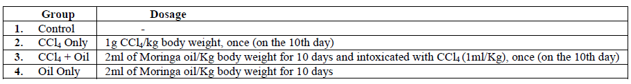
The route of administration was by oral (per orem). Twenty-four hours after the last day of treatments, the
experimental animals were sacrificed and liver tissues removed for homogenization and centrifugation. The clear
supernatant obtained was thereafter subjected to biochemical analysis.
2.4 Malondialdehyde Assay
Liver tissues were homogenized using 5mls of 0.09% normal saline in a mortar and were assayed by the method of
Beuge and Aust [12].
In practice, TBARS are expressed in terms of Malondialdehyde equivalents. MDA forms a 1:2 adduct with
thiobarbituric acid (TBA) and produces a complex which can be measured spectrophotometrically. This complex
absorbs maximally at 532nm.
2.4.1 Assay Protocol
A stock solution of trichloroacetic acid (TCA), thiobarbituric acid (TBA) and Hydrochloric acid (HCl) containing
15g TCA, 0.375g TBA and 0.25N HCl was prepared. The solution was heated mildly to assist the dissolution of
TBA. To 1ml of the tissue sample, 2ml of TCA-TBA-HCl mixture was added and heated in a boiling water bath for
15 minutes. It was cooled and then centrifuged at 3600rpm for 10 minutes. The absorbance of the supernatant was
read at 532nm against a reagent blank which contained 3ml of the TCA-TBA-HCl solution.
The malondialdehyde concentration was calculated as follows:
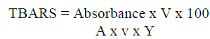
Where: Absorbance is read at 532nm; V= total volume of the reaction mixture
A= molar extinction coefficient which is 1.5x10⁵
Y= Weight of tissue; v= volume of sample used.
2.5 CATALASE ACTIVITY ASSAY
The tissues were homogenized using 0.09% normal saline solution in a mortar. The homogenate was then
centrifuged at 3600rpm for 10 minutes and the supernatant used for assay according to the method of Cohen et al.
[13].
Catalase prevents the accumulation of H2O2 by converting it to H2O and O2 according to the reaction shown below:
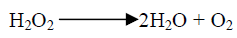
2.5.1 ASSAY PROTOCOL
0.5ml aliquot of extract homogenate was added to ice cold tubes. Reaction was started by adding 5ml of 30mM
H202. Tube contents were then mixed thoroughly by inversion. Reaction was stopped after 3 minutes with 1ml of
6M H2S04. 7ml of 0.01M potassium permanganate (KMnO4) was then added and absorbance read at 480nm within
30-60 seconds.
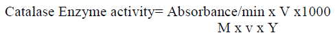
Absorbance is read at 480nm
V= total volume of the reaction mixture; A= molar extinction coefficient which is 40
Y= Weight of tissue; v= volume of sample used
2.6 Superoxide Dismutase Activity Assay
Superoxide dismutase (SOD) catalyzes the dismutation of superoxide into oxygen and hydrogen peroxide. The assay
of SOD in the liver was determined by the method of Misra and Fredovich [14].
An aliquot, 0.4mls of the diluted supernatant of the tissue homogenate was added to 5mls of 0.05M carbon buffer,
pH 10.2 to equilibrate in the spectrophotometer and the reaction started by the addition of 0.6mls of freshly prepared
0.3mM epinephrine as the substrate to the buffer supernatant mixture, which was quickly mixed by inversion.
The reference cuvette contains 5mls of the buffer, 0.6mls of the adrenaline and 0.4mls of distilled water. The
increase in absorbance at 480nm due to the adrenochrome formed was monitored every 30seconds for 120seconds.
One unit of SOD activity was given as the amount of SOD necessary to cause 50% inhibition of the oxidation of
epinephrine to adrenochrome during 60seconds.

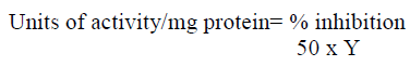
Absorbance test= Absorbance of sample
Absorbance reference= absorbance of blank; Y= weight of protein in the volume of sample used.
2.7 STATISTICAL ANALYSIS OF DATA
Using SPSS version 17 for Windows, Analysis of Variance (ANOVA) method was employed to compare the mean
differences observed among the various groups. The results are therefore presented as mean ± SD (standard
deviation) with the level of significance set at p< 0.05.
Results and Discussion
The concentration of malondialdehyde in the liver was taken as an index of hepatic lipid peroxidation induced by
toxicant CCl4, while levels of activity of both superoxide dismutase and catalase were determined as indices of
hepatic antioxidant status. The results are presented in Table 1.
Table 1. Liver MDA Concentrations, Catalase and SOD Activities among the Different Treatment Groups
CCl4 is a well-known model compound for producing chemical hepatic injury [15]. This study investigated the
protective role of Moringa oleifera seed oil against CCl4-induced hepatocellular oxidative damage. It was found that
CCl4 intoxication caused a markedly elevated hepatic MDA level (Table 1) which is an indication of increased
oxidative stress; this finding correlates with previous reports [16]. Previously, Moringa oleifera seed oil had been
suggested to be capable of reversing or inhibiting lipid peroxidation in liver cells [17]. Increased production of
intracellular ROS plays a major role in CCl4-induced hepato-cellular damage [16]. In the present study, animals preadministered
with the oil had significantly lower MDA compared with those treated only with the toxicant CCl4, an
indication of the attenuating effect of Moringa oleifera seed oil on CCl4-induced liver injury. Moringa seed oil has
been reportedly shown to contain a surplus of tocopherols for quenching free radicals and performing its other
antioxidant functions in the human body [18]. Tocopherols are natural lipid soluble antioxidants and potent free
radical scavengers present in the oil [19].
Animals pre-treated with the oil also showed significantly higher and relatively normal SOD and Catalase activities
as against those treated with only CCl4. SOD and Catalase are antioxidant enzymes considered to play crucial roles
in cellular defence against chemical- or xenobiotic-induced oxidative damage. Thus, Moringa oil acting as a dietary
(exogenous) antioxidant source, appeared to complement the antioxidant enzymes in maintaining the hepatocellular
integrity.
Conclusion
The findings from this study strongly suggest that Moringa oleifera seed oil possesses strong hepatoprotective effect
through prevention of CCl4-induced hepatocellular membrane damage. This potential of the Moringa seed oil extract
is therefore believed to be largely attributable to its natural antioxidant constituents.
Acknowledgements
The authors would like to extend their gratitude to Miss Juliet Bardi and Mr. Agu for the assistance they rendered
during this research.
References
- Doherty RE. Environmental Forensics, 2000, 1(1), 69–81.
- Oluba OM, Onyeneke EC, Ojieh GC, Idonije BO, Ojiezeh TI. Der Pharmacia Lettre, 2010, 2(4): 432-439 (https://scholarsresearchlibrary.com/archive.html)
- Sahreen S, Khan MR, and Khan RA. BMC Complementary and Alternative Medicine 2011, 11:48 https://www.biomedcentral.com/1472-6882/11/48
- Marnett LJ. Mutation research, 1999 Mar 8, 424(1-2), 83-95.
- Rahman K. Clinical Interventions in Aging, 2007, 2(2), 219–236.
- Walia A, Malan R, Saini S, Saini V, Gupta S. Der Pharmacia Sinica, 2011, 2 (5): 288-299
- Fang Y, Yang S, and Wu G. Nutrition, 2002, 18, 872– 879,
- Anwar F, Latif S, Ashraf M and Gilani AH. Phytother. Res. 2007, 21, 17–25.
- Bennett RN, Mellon FA, Foidl N, Pratt JH, DuPont MS, Perkins L, Kroon PA. Journal of Agricultural and Food Chemistry, 2003, 51, 3546-3553.
- Kumar PS, Mishra D, Ghosh G, Panda CS. International Journal of Phytomedicine, 2010, 2, 210-216. https://www.arjournals.org/ijop.html
- Directive 2010/63/EU. Official Journal of the European Union, 22 September 2010, L 276/33-79.
- Buege JA, Aust SD. Microsomal lipid, Peroxidation. In: Flesicher, S., Packer, L. (Eds.), Methods in Enzymology Vol. 52. (Academic Press, New-York, 1978) pp. 302–310.
- Cohen G, Dembiec D, Marcus J, Anal. Biochem., 1970, 34, 30-38.
- Misra HP, Fridovich I, J Biol chem., 1972, 247, 3170-3175.
- Tarantino G, Di Minno MND, Capone D, World J Gastroenterol, 2009 June 21, 15(23), 2817-2833.
- Manna P, Bhattacharyya S, Das J, Ghosh J, and Sil PC, Evidence-Based Complementary and Alternative Medicine. Volume 2011, Article ID 832805, 17 pages. doi:10.1093/ecam/neq065
- Olatosin TM, Akinduko DS, Uche CZ, The International Journal of Engineering and Science, 2013, Volume 2(11), 13-18. ISSN (e): 2319 – 1813 ISSN (p): 2319 – 1805.
- Bhatnagar AS, and Krishna AGG, Grasas Y Aceites, octubre-diciembre 2013, 64(5), 537-545. issn: 0017-3495 doi: 10.3989/gya.010613
- Christie WW. The Lipid Library. https://lipidlibrary.aocs.org/ (Accessed on 20th March 2012).


